Till Stephan
@stephan_till
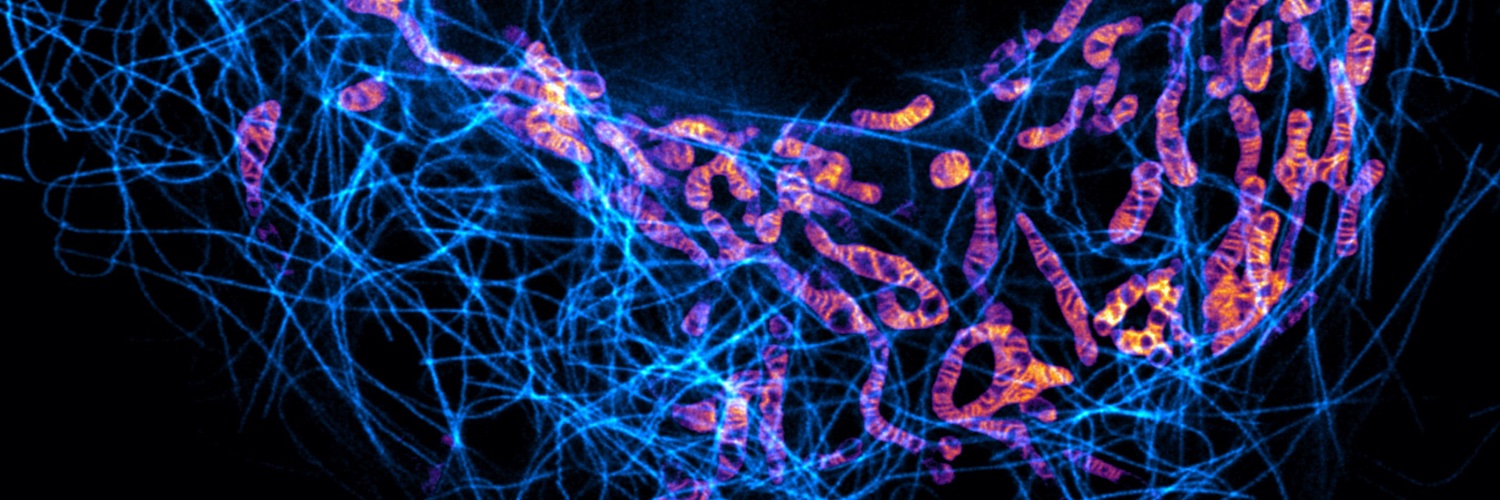
Professor at Goethe University Frankfurt. Cell biologist. Microscopist. Exploring subcellular architecture with super-resolution fluorescence microscopy.
I’m thrilled to share that I’ve been appointed as a Professor at Goethe University Frankfurt🎉 The Stephan Lab will use advanced bioimaging 🔬🧫 to explore how organelles collaborate to maintain subcellular architecture and metabolism. Find us now also on Bluesky: stephanlab.


In mitochondria, respiratory supercomplexes (blue) pump protons across crista membranes to create an electrochemical gradient, fueling adenosine triphosphate (ATP) synthase (pink) to regenerate ATP—the energy currency of life. Now, cryo–electron tomography captures these…
The final version of our work on the in-cell architecture of the mitochondrial respiratory chain is now online in @ScienceMagazine 🎉 You can find the full story here science.org/doi/10.1126/sc… @luminous_lab did a wonderful job on the cover and animation👩🎨
In mitochondria, respiratory supercomplexes (blue) pump protons across crista membranes to create an electrochemical gradient, fueling adenosine triphosphate (ATP) synthase (pink) to regenerate ATP—the energy currency of life. Now, cryo–electron tomography captures these…
Ever wondered how cells regulate the molecular traffic in and out of their nuclei? New research from Texas A&M University, supported by MINFLUX experts from the EMBL Imaging Centre, is helping uncover how molecules navigate the nuclear pore complex. embl.org/news/science-t…
In our interview with Jennifer Lippincott-Schwartz @JLS_Lab, we discuss her research career, her advice on building collaborations and learn why she is looking forward to #biologists100. Jennifer will be speaking in our emerging technologies session. journals.biologists.com/jcs/article/13…
1/ 🚨 Our new paper is online in @Nature ! As its first author, I’m enthusiastic to finally share it with you all!🎉 🧠 We discovered a mechanistic link between cellular energy metabolism and the control of the need to sleep 💤 👉 nature.com/articles/s4158… @OxfordDPAG 🧵👇
This poster is a must have for labs working on #MCS! A guide through the work of so many outstanding labs (apologies if we did not cite your work). Hang it in your office or lab, it will please your eyes and inspire your mind! Kudos to the driving force Antigoni Diokmetzidou! -LS
In their Cell Science at a Glance article, Antigoni Diokmetzidou & Luca Scorrano @LabScorrano summarise the mitochondrial contacts characterised in mammals, the mechanisms underlying their formation, and their principal functions. journals.biologists.com/jcs/article/13… #JCSMitoSI #OpenAccess
One of the most impressive 3D STED images I’ve seen in a long time!
#fluorescencefriday with centriolar rosettes in fixed RPE-1 cells, imaged confocal / with #3DSTED and #FLEXPOSURE adaptive illumination. Labeling: CP110🟥(STAR RED), SAS-6🟦(STAR ORANGE). Sample courtesy: @c_sullenberger & Jadranka Loncarek, Cancer Innovation Laboratory,…
🎉 Exciting news! The SCALE Cluster has been named a DFG Cluster of Excellence—and the Lemke Lab is proud to be part of it! This is a major step forward in understanding the architecture of life at the subcellular level.
🎉 We’re absolutely thrilled: SCALE has been selected as a DFG Excellence Cluster! A huge thank you to our incredible team—this would not have been possible without your dedication and talent. 🙌 @dfg_public #Exzellenzstrategie #SCALEcluster 🧵
Silencing mitochondrial gene expression in living cells | Science science.org/doi/10.1126/sc…. Wonderful that our paper is out now in Science. A big thanks to LD Cruz-Zaragoza and the team, @warscheidlab & @JakobsLab .
I’m thrilled to announce that my application to the Emmy Noether Program was successful! Over the next years, we’ll investigate how ER–mitochondria contact sites control the lipidome of both organelles. We’re looking for talented researchers to join us on this journey! 🎉🔬

Here come our new palette of fluorescent dyes. The BD dyes endeaves to balance glowy brightness, robust photostability, and biocompatibility. Let's boost 4D dynamic super-resolution imaging!!! nature.com/articles/s4159… @spirochrome
New piece in Journal of Cell Science on the origin and evolution of mitochondrial inner membrane structure and composition #lipidtime @Co_Biologists journals.biologists.com/jcs/article/13…
🤩
4Pi-SMS and pan-ExM reveal, in a close collaboration with the Rothman lab, a surprising tetraplex organization and self-assembly of rim golgins, which we believe can explain stack formation of the Golgi apparatus: biorxiv.org/content/10.110…
🍩🙌🏽
I’m excited to share our latest research from @musser_lab , published today in @Nature. Using two-color 3D MINFLUX, we directly visualized nuclear import and export events through transport-active nuclear pores. nature.com/articles/s4158…
🤩
In situ architecture of the human prohibitin complex nature.com/articles/s4155…
Super-res-stimulated emission depletion STED imaging of mitochondrial ultrastructure made possible in fixed cells with new fluorescent probe Image made with @LeicaMicro 📷: Jingting Chen et al @ZhixingChen2 @PKU1898 in @PNASNews ➡️: bpod.org.uk/archive/2025/3… with @DrJohnAnkers
Super-resolution microscopy of mitochondrial mRNAs Stefan Stoldt, Frederike Maass, Michael Weber, Sven Dennerlein, Peter Ilgen, Jutta Gaertner, Aysenur Canfes, Sarah V. Schweighofer, Daniel C. Jans, Peter Rehling and Stefan Jakobs biorxiv.org/content/early/…
🚨🚨Deadline approaching - last day to apply 🚨 Come and join a team of interdisciplinary microscopists as a postdoc at the University of Warwick @BMSatWarwick @warwickmed All info below⬇️⬇️⬇️
We are building a quantitative imaging team @uniofwarwick @BMSatWarwick ‼️3 postdoc positions open‼️ Join an interdisciplinary endeavor with @midbody @jitumayor_lab @SPOREMOHAN on a @wellcometrust funded project to push probe and microscopy development: warwick-careers.tal.net/vx/lang-en-GB/…