Joyce Meiring
@psychescope
Postdoc in Akhmanova lab @UniUtrecht. Cytoskeleton, 3D migration & light switchable cell biology tools | she/her. Bluesky: http://joycemeiri.bsky.social
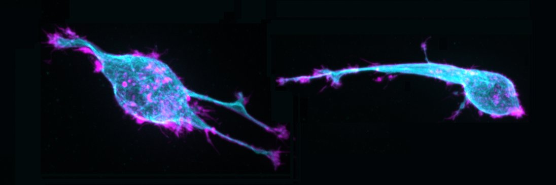
Pleased to share that the optogenetic microtubule severing tool #Optokatanin✂️ is now out in @CurrentBiology !!! authors.elsevier.com/sd/article/S09… Movie shows opto-katanin being used to locally sever microtubules in two locations (marked by blue boxes) inside a neuron.
A very elegant study by some of my colleagues on decrypting lysosomal protein trafficking! They combine synchronised transport (using the RUSH system) with APEX proximity labelling to map out the lysosomal protein trafficking pathway in neurons. 👏🏼 #POTATOMap 🥔🗺️
It's out!😭🕺First preprint as a PhD at the Farias Lab @ginny_farias where we investigated biosynthetic lysosomal membrane protein trafficking, delivery and interactome with Protein Origin, Trafficking And Targeting to Organelle Mapping (POTATOMap)🥔 biorxiv.org/content/10.110…
Radial microtubules in cyan, non-radial microtubules in yellow.
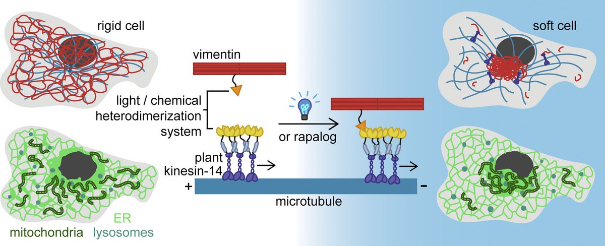
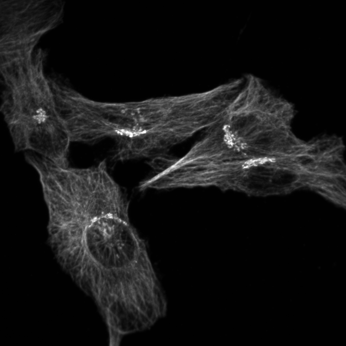
Biochemical tools to rapidly manipulate vimentin intermediate filaments by means of chemicals or light (optogenetics) 📷 Milena Pasolli & @psychescope et al Uni Utrecht, in @JCellBiol ➡️ bpod.org.uk/archive/2025/7…
Pasolli, Meiring, Akhmanova et al. (Utrecht University) describe tools to acutely relocate #vimentin intermediate filaments by inducibly coupling them to microtubule motors. hubs.la/Q03wf_F20 @psychescope @gijsjekoenderi1 #Technology #Cytoskeleton
Excited to share that our fast & reversible chemical and optogenetic #vimentin pulling tools are now published in JCB! 🥳 doi.org/10.1083/jcb.20…
Hi! I was wondering if anyone has successfully cultured happy HEK293T cells in defined serum alternatives? If so what did you use?
@ blue skies, there is bsky.app/starter-pack/t… a collection of 150 chemical biologist accounts to make it really easy to jump start your #ChemBio network. Why not at Twitter? :( no wonder people are leaving here.
.@JagerLeanne, @psirez et al. @Bijvoet_Centre show that compacted and expanded microtubule lattice spacings coexist within cells and that #microtubules recognized by a marker for stable microtubules are predominantly expanded. hubs.la/Q02SXVbD0
I'll be talking about manipulating the cytoskeleton with #photopharmaceuticals and #optogenetic tools at this Monash Advanced Microscopy webinar. Anyone can join (if you are awake at 3pm AEST)!
Caged Epothilones* now out in Angewandte: congrats Carina & all! doi.org/10.1002/anie.2… Stabilises #microtubule #cytoskeleton when and where you want. Does what you expect & nothing you don't Giving it away for bio: just mail *&more: see Fig S1! @angew_chem @photopharma_lit
Nice example of actin retrograde flow in a gigantic lamellipodium. #cytoskeleton, #actin, #microscopy, #cell-motility, #science
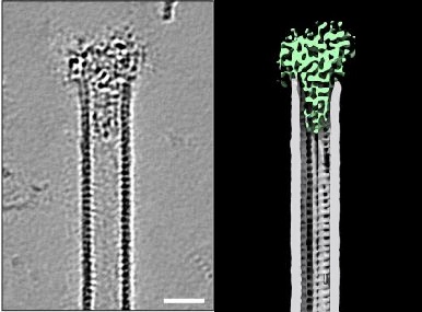
Harriet Saunders, Cyntha van de Berg, et al. show how ciliary tip proteins cooperate to make slow growing #microtubules in their new preprint! Together they form a structure that looks like a champagne cork in a #microtubule 😍 biorxiv.org/content/10.110…
Optogenetic control of the actin cytoskeleton in a neuroblastoma cell. Light is on when the white dot is visible. #microscopy #cell_control #science #cells_are_dynamic
Hi science twitter 👋 Do you test your mouse secondaries? I am looking for a reliable source of mouse secondaries for IF that don't cross react with rat, any recommendations? #Microscopy Pictured: goat anti-mouse secondary binding mouse anti-GM130 (golgi) and rat anti-tubulin
Klara Jansen, @mkiwanski, @KapiteinLukas et al. @UniUtrecht introduce StableMARK (Stable Microtubule Associated Rigor-Kinesin), a live-cell marker to visualize stable #microtubules. bit.ly/3KQroyK #Technology #Cytoskeleton