Venkatesh M☯️
@drvenkimdrd
@Radiologist ..Never ending learner, educator, #radiology #Erad #traveller , Youtuber for Shades of Radiology, Medical illustrator
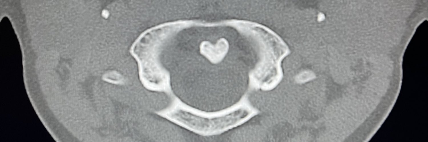
3/ 🧠 Neuroimaging: Hallmarks 🔍 CT ✅ Bilateral hyperdense thalami 🧲 MRI ✅ T1: Hyperintense bulky thalami ✅ T2: Hypointense thalami ✅ T2: Persistent WM hyperintensity ✅ Involves: subcortical U fibers, periventricular WM, internal capsule
Great case 👏
This week’s SPIN-POV: A patient with SLE shows T2 hyperintensity in the medulla, cervicomedullary junction, and spinal cord — demyelination? Infection? Don’t forget PRES with spinal cord involvement. No enhancement is seen, and there’s always a clue in the brain.
A 62-year-old man presented with a 5-day history of low back pain and a 1-day history of lethargy. Physical examination was notable for tenderness over the lumbar spine and a normal neurologic examination. Radiographs of the lumbar spine showed gas within L4. Computed tomography…
*"Proud to be part of Radiopaedia Conference! 🎉* I'm thrilled to share that our poster, Neurovascualr conflicts - Close encounters , was a huge success at the Radiopaedia conference! 🌟 It was an incredible experience presenting our work to a global audience of Radiology

Small Bowel Enteritis due to ACE Inhibitors (Lisinopril) From "Abdominal Pain in the ER: GI Pathology - Part 3" View the full lecture here: ctisus.com/media/2024/03/…
📆 Just 4 days to go! Don’t miss the @Radiopaedia 2025 Conference 🩻 Join thousands worldwide for expert-led, case-based teaching you won’t find anywhere else! 🗓️ July 21–25 👩🏻💻Conference schedule: drive.google.com/file/d/1CbdZz3… 📝 Register: radiopaedia.org/courses/radiop…


Never heard of this rare entity Thanks for sharing 👍
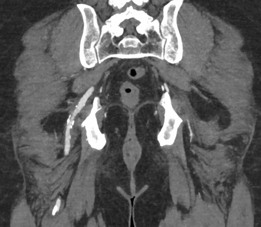
A weightlifter developed intense back and abdominal pain during competition. An ultrasound led to CT urography. Have you even heard of this diagnosis before? Images in 𝑅𝑎𝑑𝑖𝑜𝑙𝑜𝑔𝑦
A weightlifter developed intense back and abdominal pain during competition. An ultrasound led to CT urography. Have you even heard of this diagnosis before? Images in 𝑅𝑎𝑑𝑖𝑜𝑙𝑜𝑔𝑦
TEACHING COD: super subtle case of endometriosis involving the rectum; spiculated T2 hypointense mass with foci of T1 hyperintense signal#yaleradedu #yaleradiology #FOAMrad #FOAMed #radres #medstudent #radiology @RadDiscord #MedTwitter @YaleRadRes
Context: intraventricular pediatric mass 🧠 One peak to rule them all: choline. No NAA. No creatine. 📉 Neuron-free profile. 🎯 Highly suggestive of choroid plexus tumor. #MRI #PediatricNeuro

Great team work 👏👏 Central vessel Convolute 🤩
#JustPublished: Novel anatomical observation: Prominent nutrient vessels in the ilium bone are very common, and mostly form a distinct, recognizable branching pattern, the Central Vessel Convolute (CVC). 👉Read the article for free: doi.org/10.1007/s00256… @derbalgrist #MSKrad
Join the webinar this coming Sunday on Improving fracture detection on plain radiography 😎 Details below 👇🏻👇🏻
June 8 - Sunday International Webinar#857 Improving Fracture detection - Strategies on Plain Radiography - Dr. Venkatesh Manchikanti, Specialist Radiologist, UAE Join here: us02web.zoom.us/j/85666220065?… Webinar ID: 856 6622 0065 | Passcode: REFCAFE
#FOAMrad #FOAMed #radres #radtech #medstudent #radquarters
I’m pleased to share with you our new publication in RadioGraphics on the imaging features of adenomyosis, including typical and atypical imaging appearances at US and MRI @RSNA @RadioGraphics @cookyscan1
#ASHNRCOTW #294: ANSWER. Thx Dr. Warren 4 case! #ASHNR25 @callyrobs @DShatzkes @CDP_Rad @rhwiggins @nakoontz @KRileyMD @CMGlastonbury @tabby_kennedy @PhilipRChapman1 @cmtomblinson @amyfjuliano @bpoliceni @MohitTCCNeuro @AshokSrini15 @KatieTraylorD @CynXinWu
"Snoopy sign" of pericardial agenesis 🔹Cardiac levoposition 🔹Lengthening and flattening of the border of LV 🔹Lucency separating the LV and left hemidiaphragm 🔹Lucency separating the pulmonary artery and aorta (interposed lung) 🔹 Loss of the right heart border PC: REF/CMC
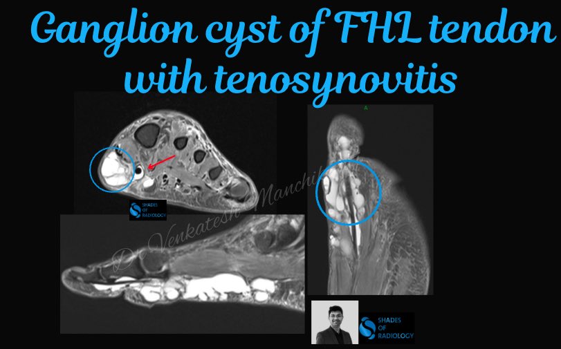
FHL Ganglion cyst - Rare case presentation 👇🏻👇🏻 @mskradiologyuk @PrimeFellowship

🟢 Groin pain in soccer players: anatomy, clinical presentation, biomechanics, pathology and imaging findings ⚽ Read more 👉 rdcu.be/efECx #SkeletalRadiology #Soccer #SportsMedicine