Tapas Kumar Sahu
@doc_tks_rad
Radiologist. Fellowship in Oncoimaging. Assistant Professor @ KIMS, Bhubaneswar, India #FOAMrad #Medtwitter #Radiology
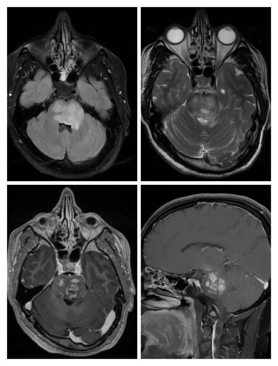
Chronic lymphocytic inflammation with pontine perivascular enhancement responsive to steroids (CLIPPERS) As the name suggests characterized by multiple punctate, patchy and linear areas of enhancement in the pons and typically respond well to steroids✅ #FOAMrad #neurotwitter
🧠⚙️ #SpinTweetorialWednesday Let’s decode FAHN – Fatty Acid Hydroxylase-Associated Neurodegeneration, aka SPG35. An NBIA subtype with spasticity, ataxia, dystonia & brain iron accumulation—but no “eye of the tiger”! 🧵👇
This week’s SPIN-POV: An “L”-shaped thalamic hyperintensity on DWI/T2 points to partial prolonged HIE in term neonates—exaggerated by hypoglycemia but uncommon in isolation. In preterms, it is associated with parieto-occipital PVL, helping differentiate it from metabolic causes.
This week’s SPIN-POV: New neurological symptoms in a patient with known CVT? Always rule out a dural AV fistula. Symptoms occur due to venous hypertension. Often subtle, they may show as flow voids on T2 and prominent serpiginous vessels on SWI, just like in this case.
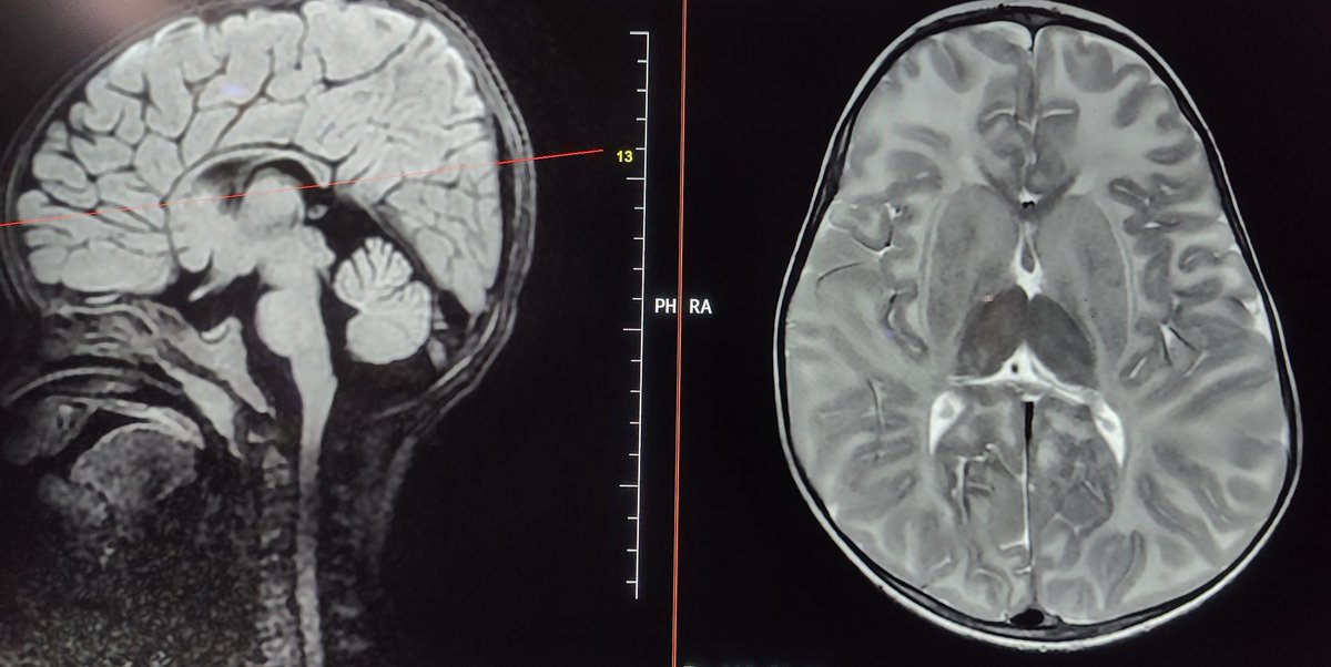
🧠✨ Ever seen T1 bright, T2 dark thalami on MRI in an infant? That thalamic whisper could be Sandhoff disease — a devastating form of GM2 gangliosidosis. This #SpinTweetorial dives into the clinical, imaging & genetic clues. Let’s go 🧵👇 #NeuroRad #PedsNeuroRad
SPIN Tweetorial Wednesday: Tay–Sachs Disease on MRI Tay–Sachs disease is a lysosomal storage disorder under the GM2 gangliosidosis umbrella. Today, we’re decoding its characteristic MRI fingerprints. Let’s SPIN! 🧵👇
#RadRes: Anterior osteophyte fx. can be tricky in a degenerative spine: ⤵️ Left = acute Right = chronic ✅ Prevertebral effusion & absent cortication along the "lucency" although are super helpful... #NeuroRad #Trauma #Spine
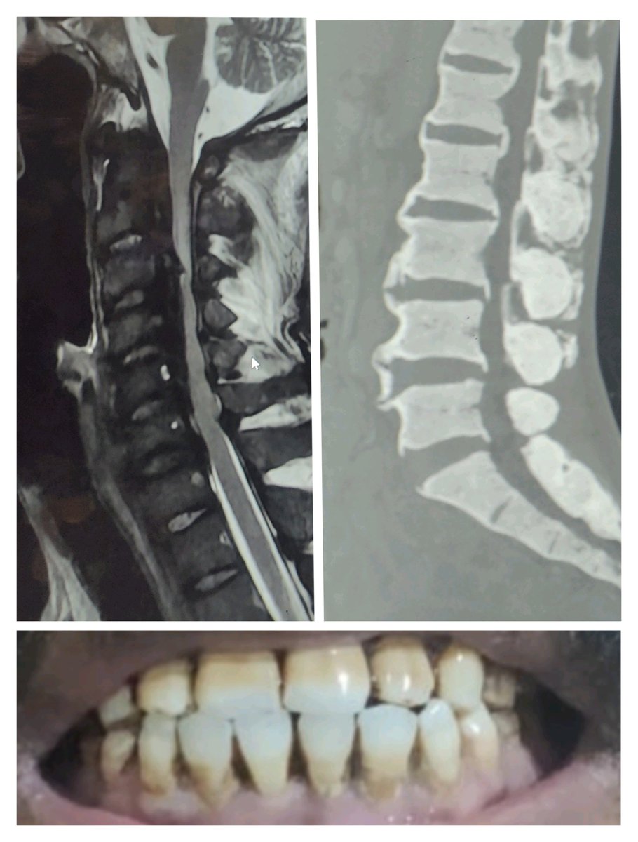
SKELETAL FLUOROSIS 🔷Diffuse osteosclerosis / T1 & T2 hypointense marrow on MRI 🔷OPLL & ossification/calcification of other paraspinal ligaments, bony excrescence ➡️spinal canal/foraminal compression➡️radiculomyelopathy ✅Look for dental fluorosis ✅Lab tests: Urine fluoride
Ethmoid cells are separated into anterior and posterior ethmoid by the basal lamella, the lateral attachment of the middle turbinate. ESHNR webinar “Paranasal Sinus Anatomy and Variants” by @drSurjthVattoth from Little Rock/US @ASHNRSociety @BSHNI_UK @ESNRad @myESR
7 Sci-Hub Alternative Websites Paper you need to ask for payment & can't use sci-hub? You don't have to pay to read academic papers. These are 7 sci-hub alternative websites to download papers for free.
How to Use AI to Read 10 Research Papers in 30 Minutes Tired of spending hours reading academic papers? 😩 Here’s how to use AI to speed up your reading and actually understand what matters 👇
For Neonatal CNS infections...Neurosonography is a great tool!
Do you routinely use cranial ultrasound? We do — and for good reason. As a screening and monitoring tool, it's incredibly effective in selected neonatal cases. This paper is worth reading — findings can be trickier than they seem 🧠📖 #PedsRad 🟢🔓pmc.ncbi.nlm.nih.gov/articles/PMC12…
SANDHOFF DISEASE- genetically proven 🔷GM2 gangliosidosis 🔷Marked T1/T2 shortening of b/l thalami 🔷Diffuse WM hyperintensity ➡️ dysmyelination or demyelination 🔷Basal ganglia (caudate & putamen) also involved 🔷 J-shaped Sella (not a specific finding) #neurotwitter #FOAMRAD
Rabies Encephalitis!!
🔷What is the most likely diagnosis in this 70 y/o F who lives with feral cats presenting w/ vomiting, diarrhea, leukocytosis, fever for 3 days and progressive decline in level of consciousness? 🔷CSF: initially normal, repeat a few days later ⬆️ WBC (lymphocyte predominant), ⬆️…
Up to 1/3rd of patients develop fatty liver after Whipple surgery, with severe cases like NASH or cirrhosis linked to malnutrition, pancreatic insufficiency, and metabolic disruptions. Risk Factors: Female gender, pancreatic cancer, rapid weight loss &low panc enzyme output.
🔷Imaging hints: ▶️Cortical sulci do not reach the inner table of the skull (yellow arrows) and are partially effaced ▶️Mass effect ▶️Apparent thickened cortex ▶️If contrast was given, displaced cortical veins between the collection and the cortex may be helpful
Today was a routine day in OPD. It is not always CT/MR, sometimes X rays bark the Dx. 50M. 6M pain around L iliac crest. No local tenderness. Rx: NSAIDS + steroids, no response. We got X ray pelvis. Seen this after a long time. #MedTwitter #neurotwitter #radtwitter #radres
Hx: "seizures" Looks up vitals: SBP 220 Dx: HBE (Hypertensive Brainstem Encephalopathy) ➡️ Pontine predominant variant of "PRES" No differential diagnosis needed! 😉
Rule of thumb for detecting M2 MCA vessel occlusion on CTA: ⤵️ ✅ Superior M2 division = territory predominantly ant. to sylvian fissure. ✅ Inferior M2 division = territory predominantly post. to sylvian fissure. Real time saver on those busy call nights 😉!
Quadrigeminal cistern lipoma ✅Intracranial lipomas show SWI blooming- but not due to blood products or Ca²+ ✅SWI, highly sensitive to magnetic susceptibility differences➡️ fat can create local field inhomogeneities➡️ signal dropout/blooming at the edges (India ink artifact)!

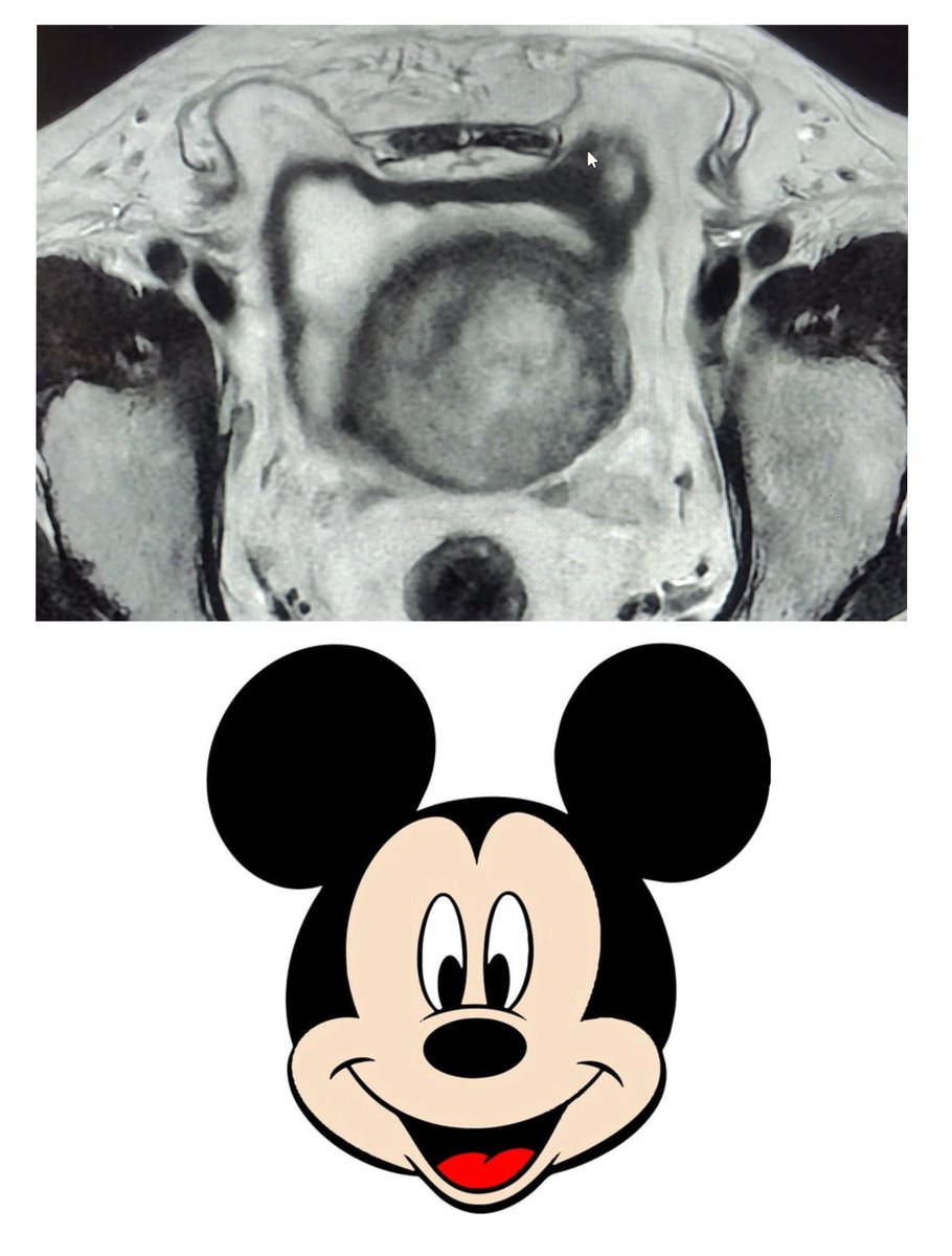
Bilateral Inguinal Hernia (direct) containing omental fat & anterolateral UB wall as contents- "Mickey Mouse sign" 🐭 on MRI Ignore the partial volume of gross prostatomegaly!! #FOAMrad #MickeyMouse