
Reto Sutter
@SutterBalgrist
Professor and Chief of Radiology at Balgrist University Hospital @derbalgrist University of Zurich @UZH_ch; all things musculoskeletal #mskrad; tweets my own
How to best diagnose #Conjoint #NerveRoots on spine MRI? Read it here for free 👉 doi.org/10.1007/s00256… The coronal #STIR sequence greatly improved sensitivity and inter-reader agreement for conjoint lumbar nerve root (CLNR) detection on MRI. @skeletaljournal @derbalgrist

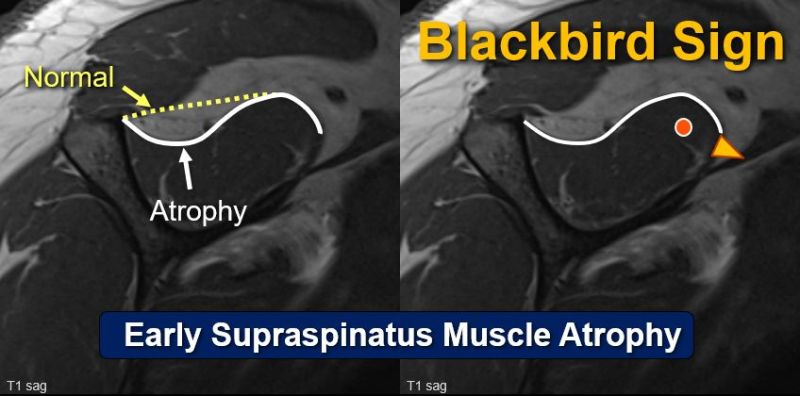
The #BlackbirdSign: Out now in print & #OpenAccess 👉link.springer.com/article/10.100… This sign can identify #EarlyAtrophy of the #Supraspinatus muscle, which is an important risk factor for re-tears after tendon repair of the shoulder. @FeuerriegelG @derbalgrist @UZH_en @EurRadiology
The findings support expanded use of photon-counting detector CT for lumbar spine evaluation. ajronline.org/doi/10.2214/aj…
#JustPublished: Unicompartmental Knee Arthroplasty (#UKA): What are the Radiographic Predictors for #Conversion to Total Knee Arthroplasty (#TKA)? 👉 Access the full article here (#OpenAccess): doi.org/10.1016/j.ejra… @derbalgrist @BalgristCampus #MSKrad

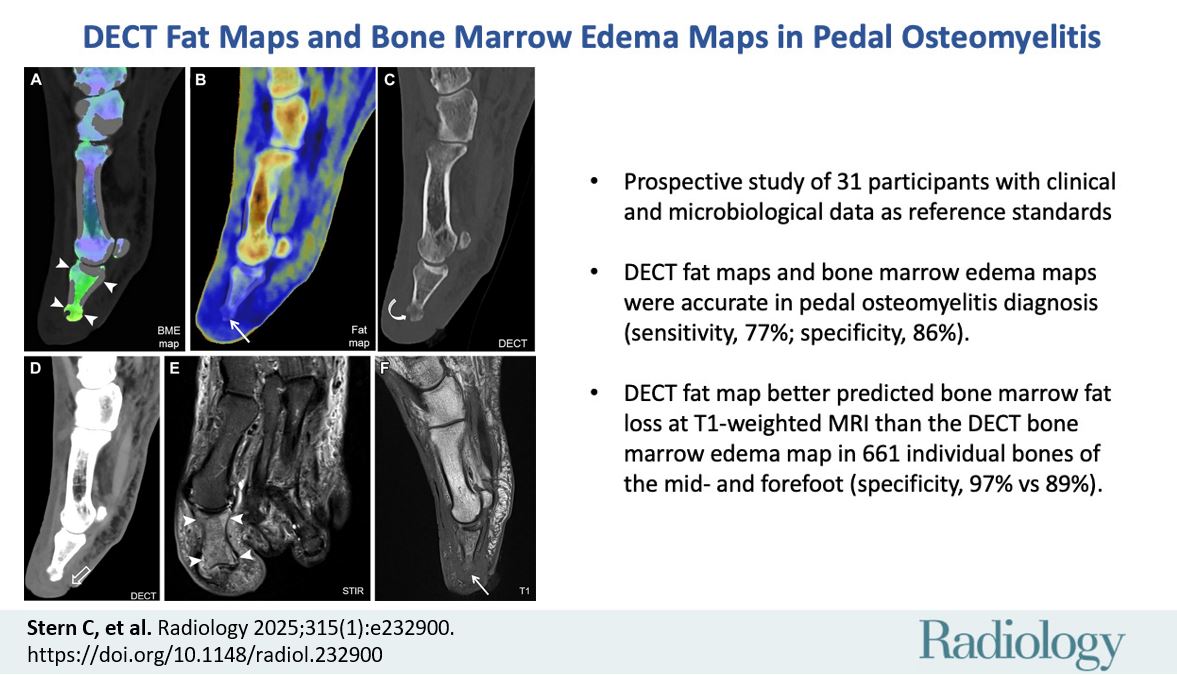
#JustPublished: Novel DECT Fat Maps for diagnosing Osteomyelitis. Dual Energy CT Fat Maps can be used together with Bone Marrow Edema Maps for detecting #Osteomyelitis 👉Read the article here: pubs.rsna.org/doi/10.1148/ra… @derbalgrist @BalgristCampus @radiology_rsna @RSNA #MSKrad
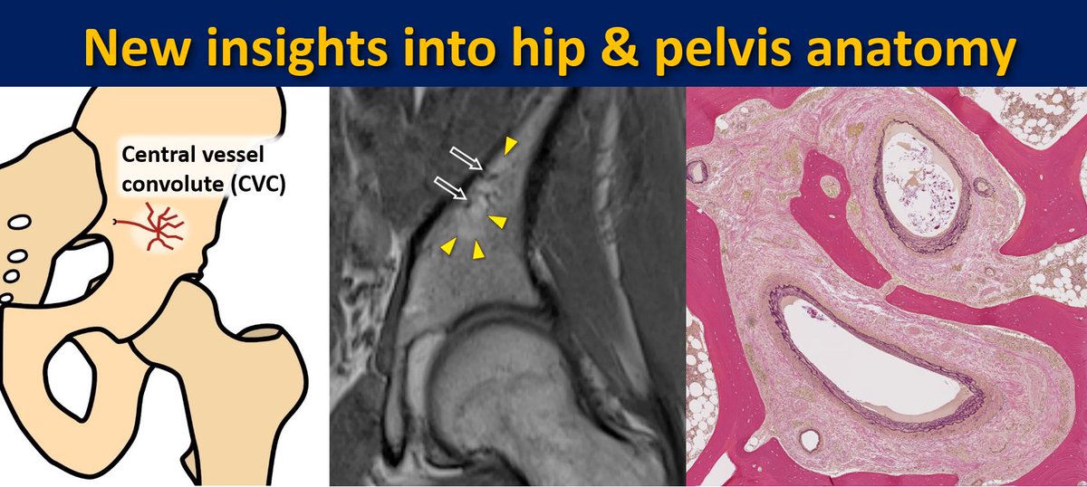
Great team work 👏👏 Central vessel Convolute 🤩
#JustPublished: Novel anatomical observation: Prominent nutrient vessels in the ilium bone are very common, and mostly form a distinct, recognizable branching pattern, the Central Vessel Convolute (CVC). 👉Read the article for free: doi.org/10.1007/s00256… @derbalgrist #MSKrad
#JustPublished: Novel anatomical observation: Prominent nutrient vessels in the ilium bone are very common, and mostly form a distinct, recognizable branching pattern, the Central Vessel Convolute (CVC). 👉Read the article for free: doi.org/10.1007/s00256… @derbalgrist #MSKrad
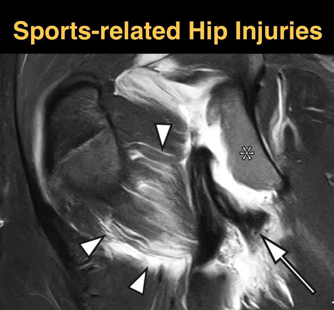
Happy to be part of the special #SportsImaging issue of @SMR_SeminarsMSK for #ESSR2025! Read our article on Sports-related Hip Injuries here: doi.org/10.1055/s-0045… #MSKrad @derbalgrist @UZH_ch
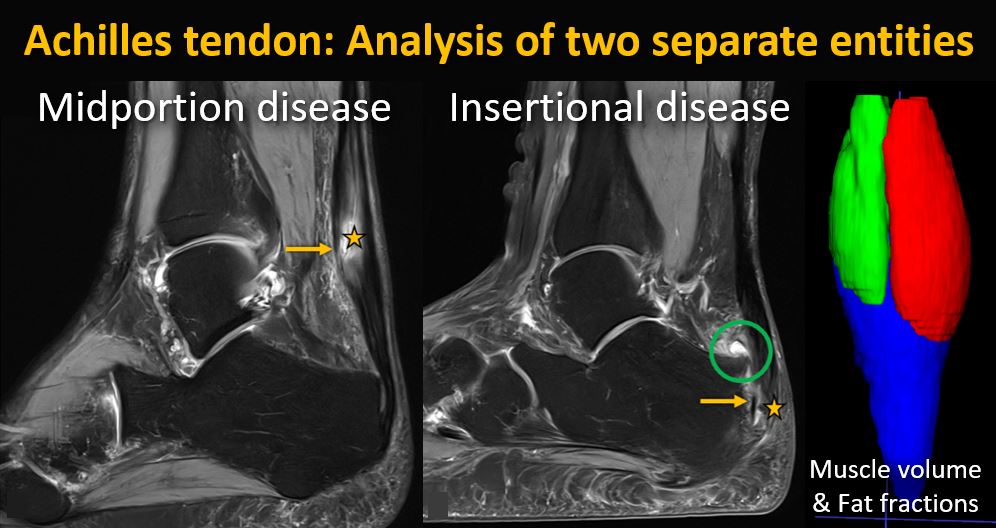
Now out in print and #OpenAccess here 👉link.springer.com/article/10.100… #Achilles tendon disease comes in two entities: #Midportion disease: More severe tendon thickening & complete tears #Insertional disease: Partial tears more frequent @derbalgrist @skeletaljournal @sophiasamira92

Check out our latest article in Radiology here 👉doi.org/10.1148/radiol… We discuss the latest MRI techniques for assessing the #shoulder and rotator cuff muscles and explore advanced methods for quantifying muscle degeneration @FeuerriegelG @derbalgrist @radiology_rsna #MSKrad

How much can MRI reveal about rotator cuff health? 💪🧲This study by @FeuerriegelG, @SutterBalgrist and team shows how qualitative and quantitative MRI assess fatty degeneration and muscle atrophy, offering key clues for outcomes. #MSK #Radiology #MRI pubs.rsna.org/doi/10.1148/ra…
#Reference: Feuerriegel GC, Fritz B, Marth AA, Sommer S, Wieser K, Sutter R. Assessment of the Rotator Cuff Muscles: State-of-the-Art MRI and Clinical Implications. Radiology. 2025 May;315(2):e242131. doi: 10.1148/radiol.242131.
➡️ This review explores both qualitative and advanced quantitative MRI techniques for assessing RC muscle fatty degeneration and atrophy, as well as the clinical importance of intramuscular fat quantification in surgical decision making and outcome prediction.
Do you know how the #RCmuscle changes after tendon tear and how it is assessed on MRI? 👉Read our recent review in @radiology_rsna : doi.org/10.1148/radiol… @SutterBalgrist @derbalgrist @BalgristCampus #RadInTraining @RITEditor
Don’t Miss it! @DrIanWeissman @Vilavaite @agtenc @AChhabraMD @erinalaia @BCOrthopods @balvarezdsierra @brucebforster @dblankenbaker2 @BCRadSoc @MarceloBordalo @carlespedret @tatcantarelli @canadaradwomen @DrMayuran @msk_munoz @MskSerme @MSKMarSol @mairaleite_msk @SutterBalgrist
Want to receive the event zoom links and the 7 day links to view the recordings? Sign up at bonesquad.ca @RolaHusain For charitable donations: give.ubc.ca/musculoskeleta…
While #MRI performs best for osteomyelitis, #DECT with #BME and fat maps is a viable diagnostic alternative to detect osteomyelitis in patients who cannot undergo MRI. 👇 pubs.rsna.org/doi/10.1148/ra… @DrLindaMoy @RadiologyEditor @VChernyakMD @RITEditor #RadInTraining
For patients with #osteomyelitis who cannot undergo #MRI, can #DECT (Dual Energy CT) be the solution? Check it in this #Tweetorial! #RadInTraining 👉pubs.rsna.org/doi/10.1148/ra… @DrLindaMoy @RadiologyEditor @VChernyakMD @RITEditor
#Achilles tendon disease comes in two entities: Read our analysis for free👉link.springer.com/article/10.100… #Midportion disease: More severe tendon thickening & complete tears #Insertional disease: Partial tears more frequent @derbalgrist @skeletaljournal @sophiasamira92 #MSKrad
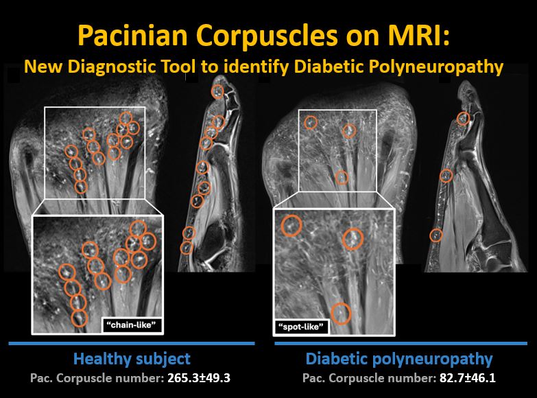
New #MRI study about #PacinianCorpuscles in diabetic patients with polyneuropathy: Read it for free 👉insightsimaging.springeropen.com/articles/10.11… 🔵Reduced number & altered pattern of corpuscles may predict diabetic polyneuropathy @derbalgrist @InsightsImaging @sophiasamira92 #MSKrad #OpenAccess
Reverse Shoulder #Arthroplasty: What are normal and abnormal CT findings? 👉 #OpenAccess Article: link.springer.com/article/10.100… Many seemingly abnormal #CT findings can be seen in both patient groups. #Dislocation was the only finding significantly associated with revision surgery.
