Optical Nanoscopy
@OptNanoscopy
* Thoughts on #imaging, #microscopy and optical #nanoscopy / #superresolution microscopy! * Tweets by @Ulrike_Boehm - Enjoy!
🔬 Are you looking to keep up with emerging trends in optics and photonics? Check out these tips to keep your knowledge and expertise on the forefront of optics and photonics! 😀 👉 ulrikeboehm.org/keeping-up-wit…
Imaging scientist @juliafrodri is part of a collaborative team developing + implementing novel sample preparation workflows, imaging acquisition & correlated software for multimodal imaging across scales
Just scratching the surface of what #Minflux can really do in tissue…:
#MINFLUX fluorescence nanoscopy in biological tissue New preprint by T. Moosmayer, K.A. Kiszka et al. including PSD95 relative to the spine morphology (example below), presynaptic VGlut clustering and AMPAR clustering at the post-synapse. Check it out at biorxiv.org/content/10.110…
🔬 Conducting experiments in a vibration-free environment is crucial, especially for optical systems that require precise alignment. Learn more about vibration sources & how to mitigate them 😀👉 ulrikeboehm.org/sources-of-vib…
Say hi to Carl, the very first mini version of our founding father! 👋 Born on this day 208 years ago, Carl Zeiss spent most of his life in #Jena, defining the history of optics. Now, the limited edition #PLAYMOBIL figure can keep his spirit alive. Happy birthday, Carl Zeiss! 🎉
A cell photographed through a microscope. DNA in the nucleus, the Golgi apparatus, and actin filaments are shown. #CellBiology
It is time to celebrate 🍻🍻🥮🥮 Congratulations @andrea_bucci13, @GTortarolo, @MarcusOHeld, Luca Bega, @EleonoraSPerego, @FCRayleigh, @BozzoniIrene, @EliSlenders Thanks @IITalk @ERC_Research @CSP_live
Optics, such as lenses & mirrors, play a vital role in various fields, including #microscopy, & laser systems. To ensure optimal performance of these optical components, it is crucial to handle and clean them correctly. Here are some recommendations: 😀👉 ulrikeboehm.org/cleaning-optic…
Fun chat with Peter O’Toole @YorkBioimaging happy to be part of the #Microscopists podcast serie
Chill out with Ilaria Testa (@karolinskainst) whilst she chats about: - The challenges and triumphs of grant writing - Balancing family life with groundbreaking science - Her love for Magritte's paintings because - Her lab's award-winning cake shaped like a neuron! See/hear:…
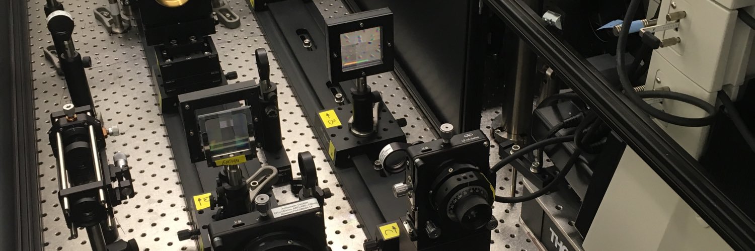
🔬 Building a new optical setup? Here are some important points to keep in mind, drawing from my experience working in optical laboratories... @SPIEtweets @euro @europeanoptics @OpticaWorldwide #optics #photonics 😀👉 ulrikeboehm.org/tips-for-build…
Thank you for visiting @HHMIJanelia, @BalzarottiFran! You will be able to watch Francisco's fantastic EMBL & Janelia bioimaging seminar on our YouTube channel soon: youtube.com/playlist?list=… @EMBLHeidelberg @markkitti @kaydanych @Prevedel_Lab @IMPvienna
How many synapses can you detect in a single Field of View? The answer is many😊. Here we have a 7plex image with 450 Synapses imaged with SUM-PAINT @Jungmannlab, that I am excited to share with you. Happy #FluorescenceFriday
#FluorescenceFriday Also a good time to mention my lab is still recruiting graduate students!
Here are some neural fireworks for 4th of July! A 3D reconstruction of a pair of optogenetically triggered spikes in a CA1 pyramidal cell, acute slice. Stim to blue branch. With Liam Paninski's lab, supported by @cziscience. biorxiv.org/content/10.110…
#FluorescenceFriday #cellbiology #bioart A neutrophilic HL-60 cell exhibits both sheet- and rosette-like dynamic pseudopods as it migrates in 3D through a collagen mesh. Here, the cell center is marked in yellow, and each pseudopod is marked with an orange ball of size…
(1/2)🌟In our speakers line-up, we introduce @S_LevequeFort, @CNRS Researcher Director at @ISMOlab_Orsayin @UnivParisSaclay. With a PhD in Optical Lab @ESPCI_Paris and postdoc at @imperialcollege, cofounded @abbelight. She is recipient of Irène Joliot Curie prize.
(1/2)Today we announce our speaker @BalzarottiFran With a PhD in Optics and Plasmonics in @UBAonline, he postdoc in @MPIbplead lead by the @NobelPrize laureate @Stefan_W_Hell🎖️and working with #superresolutionmicroscopy he developed #MINFLUX 📅Register and meet him at #SMLMS2024
So grateful to @RoyalMicroSoc and truly honored for the recognition
Many congratulations to Ilaria Testa @IlariaTesta4 @KTHuniversity who is the latest winner of the RMS Award for Light Microscopy! Read more about all our Section Award-winners: rms.org.uk/resource/rms-s… #LightMicroscopy
We're currently looking for an 👉 intern or Master's student 👈 to work on a research project that delves into the potential of deep learning-based methods for #microscopy 🤖💻🔬 #zeiss #teamzeiss Interested? We can't wait to hear from you 👉 zeissgroup.wd3.myworkdayjobs.com/External/job/O…
Now on @biorxivpreprint - I'm delighted that the collaboration between @GalbraithLab, @sciencethecat, & myself has finally come to fruition. Learn more about how compartmentalized cytoplasmic flows direct protein transport to the cell’s leading edge...😀👉 biorxiv.org/content/10.110…