Dylan Burnette
@MAG2ART
Cell biologist studying how a heart grows and dies; also Blebbisomes. Associate Professor at Vanderbilt. Married to @gillianhoo.
I am excited to finally be able to share with you our work reporting the first Extracellular Vesicle with a personality! This video does not show a cell, it is a Blebbisome! #CellBiology nature.com/articles/s4155…
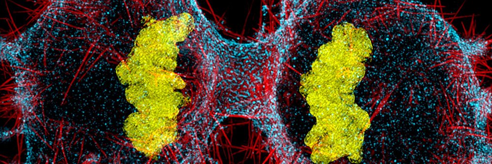
Sarcomeres within heart muscle cells produce the forces driving your heart beat. This electron micrograph shows sarcomeres in a cell from a mouse's heart. Turquoise shows the core of each sarcomere. Pink shows mitochondria, which supply the fuel: ATP #CardioCellBiology
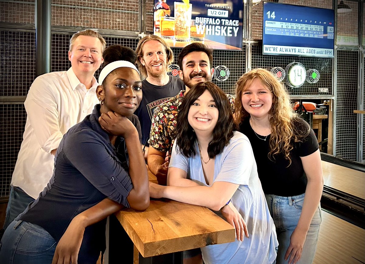
Burnette Lab outing to Pins Mechanical Nashville. Sadly, Burnette Lab 2.0 is coming to an end but Burnette Lab 3.0 is booting up! #CellBiology
Happy #microscopymonday. Today we have the back cover of our latest Thematics #issue on the CYTOSKELETON OF #MICROBES in #CYTOSKELETON. You can find the issue here: onlinelibrary.wiley.com/toc/19493592/2… . Enjoy!
I asked ChatGPT to draw a blebbisome. The first extracellular vesicle with a personality indeed :-) #CellBiology nature.com/articles/s4155…

Mag2Art.com: Activewear and canvas prints featuring photographs of mitochondria taken through a microscope. Why do these exist? I was bored. This link takes you to all of the products that turned out well: mag2art.com/collections/al…

Happy #FourthofJuly! 🇺🇸 Whether you're spending time in the lab or enjoying fireworks with friends and family, enjoy this red (nucleus), white (Golgi apparatus), and blue (actin filaments) crawling cell videoed through a microscope. 📸: Dylan Burnette @MAG2ART
Large extracellular vesicles called "migrasomes" forming from thin retraction fibers left behind by a crawling cell. Method DIC microscopy. 5 micron scale bar. 120 min. #CellBiology
A zebrafish embryo photographed through a microscope. Actin filaments are shown. You may have to zoom in to see the details...... #CellBiology

A heart muscle cell (cardiac myocyte) photographed through a microscope by PhD candidate @EmmaKoory. #CellBiology

Are my basic scientists still here? Haven’t done #FluorescenceFriday in quite a bit. Feeling sad about the state of science in the US? Go stare at some fibroblast actin networks (shown here) in a microscope until your eyes bleed! It works!
The leading edge of a crawling cell videoed through a microscope. Watch the actin cytoskeleton in motion as it drives membrane protrusion and cell movement. It’s like watching the cell think with its feet. #CellBiology
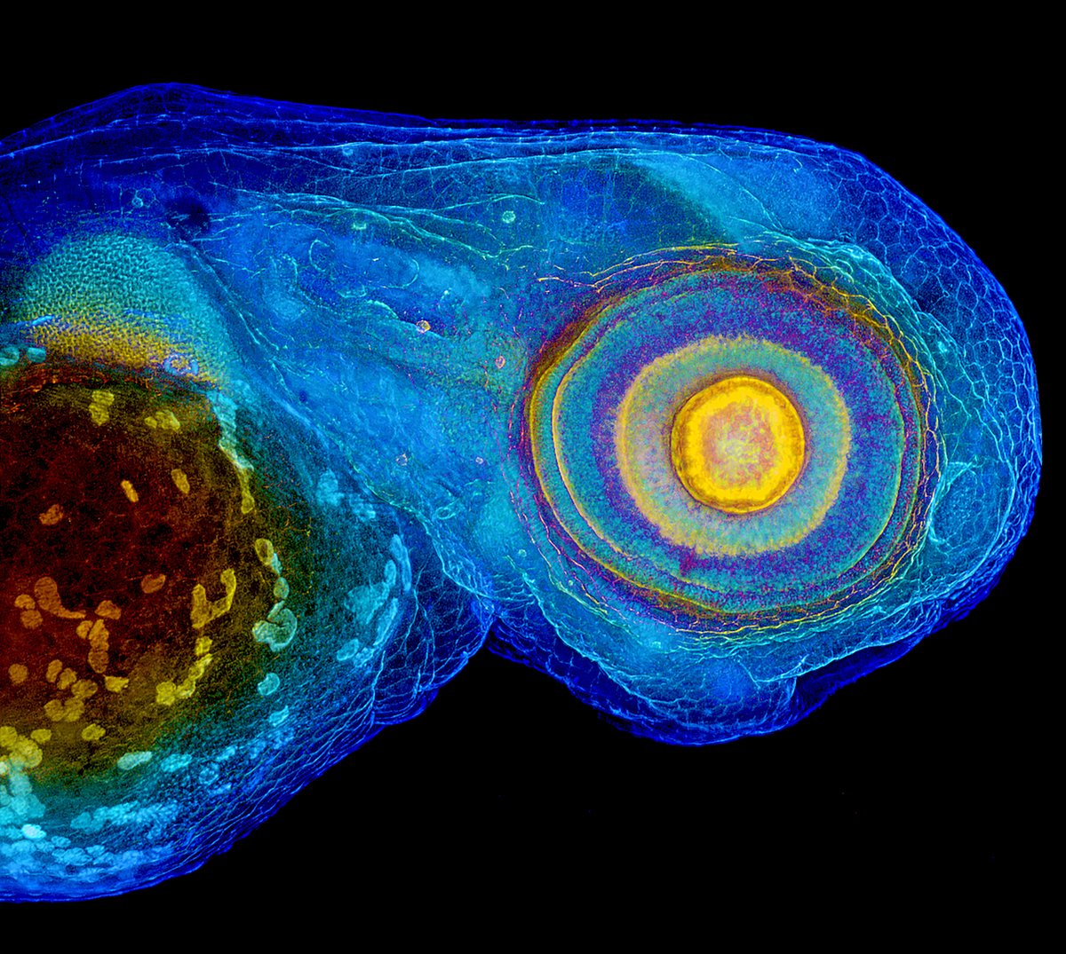
The head of a zebrafish embryo (72 hpf) photographed through a microscope. Yes, its eye follows you..... #CellBiology
An epithelial cell videoed through a microscope. #CellBiology
Burnette lab graduate student, Zach Sanchez, won the Steve Hann Outstanding Graduate Student Award! He then gave a fun talk on blebbisomes! You can read about his work here: nature.com/articles/s4155… @VUBasicSciences @EntoSanchez


