Eduard Unterauer
@EduardUnterauer
Physicist | PhD Student @JungmannLab @MPI_Biochem @LMU_Muenchen Super-resolution microscopy 🔬🧬 Spatial-omics technology 👨🎨🌈 Neuroscience Applications 🧠
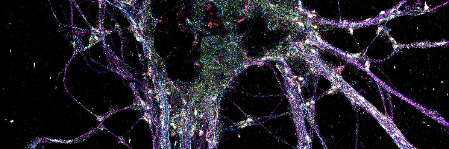
Looking at this beautifully detailed neuron, stained for 9 different proteins using SUM-PAINT, feels like stepping into one of David S. Goodsell's paintings illustrating the crowded cellular environment. #FluorescenceFriday #phdlife @JungmannLab
HUGE CONGRATS to Joschka Hellmeier and Sebastian Strauss for developing and leading this amazing work!!! Huge congrats also to all coauthors @l_masu, @shuhan_paint, @rafalkowalew, @EduardUnterauer for the great teamwork!! 7/7
Are you struggling to achieve high multiplexing at single-protein resolution and the methods section of publications leaves you puzzled? Look no further! We present our SUM-PAINT spatial proteomic imaging protocol, now published in STAR Protocols. star-protocols.cell.com/protocols/4066
New protein labelling SUM-PAINT enables high throughput, high res, multiplex imaging 📷: @EduardUnterauer, Sayedali Shetab Boushehri & Kristina Jevdokimenko et al @euforna @uniGoettingen @JungmannLab @MPI_Biochem in @CellCellPress ➡️: bpod.org.uk/archive/2024/1… with @AntDLewis
At the end of the PhD journey, you realize the goal was actually only part of the deal. It’s the people you get to know along the way and the experiences you share that shape you to become a wiser and happier human being. Thanks for the great time @JungmannLab

We are live at the @LMU_Muenchen Physics Science Fair! Come to our poster to learn about what we do at the @JungmannLab! We have several research projects for students at Bachelor and Master level!
Whipped up a DNA-themed cake for the @JungmannLab - sweet science at its finest! 🍰🔬 #PhDlife
Do you know what I love most about my PhD @JungmannLab? Actually seeing the things I learned from biology textbooks come to life🪄. Check out this DNA-PAINT image of the Golgi, showing both cis (GM130) and trans (TGN38) faces together with peroxisomes. Happy #FluorescenceFriday😊
What makes for a great DNA-PAINT experiment? How does the fluorescent dye affect the image quality? And how can we use this to extract the most out of our samples? Check out our paper in @NatureMethods to see what state-of-the-art DNA-PAINT can do for you! doi.org/10.1038/s41592…
Very proud to be part of this exciting work 😊. It's a great step forward in enabling highest resolution DNA-PAINT for everyone 🌈
What is a great DNA-PAINT experiment? How does the fluorescent dye affect the image quality? And how can we extract the most information from our samples? Check out our paper in @naturemethods to see what state-of-the-art DNA-PAINT can do for you! nature.com/articles/s4159…
Ready to present the SUM-PAINT story at the single molecule approaches to biology @GordonConf in beautiful Maine 😊. If you are interested, stop by Wednesday or Thursday 🙂

Excited to attend my first ever conference on single molecule approaches to biology by @GordonConf in beautiful Maine! Caught a fun 'Where's Waldo' moment in the hotel lobby - can you spot Ralf Jungmann and Luciano Masullo waving at me in the background? 🔍 @JungmannLab @l_masu
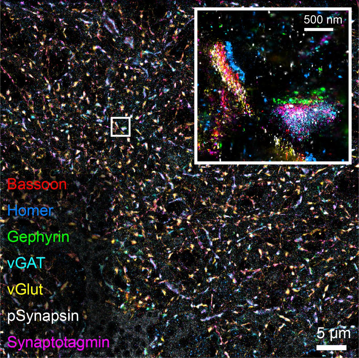
How many synapses can you detect in a single Field of View? The answer is many😊. Here we have a 7plex image with 450 Synapses imaged with SUM-PAINT @Jungmannlab, that I am excited to share with you. Happy #FluorescenceFriday
Happy Friday! To wrap up another exciting week of learning highly multiplexed DNA-PAINT in the @JungmannLab, here's a nice #cellfie of a rat hippocampal neuron stained for synaptic vesicle markers (Vamp2, VGlut1 and synaptotagmin), clathrin, neurofilament and peroxisomes.
Excited to share that I am starting a lab focused on deep learning-based protein design, biophysics and fundamental biology at @MPI_Biochem and @GeneCenter_LMU in Munich with the Emmy Noether Programme this summer. Join us as a PhD or postdoc. More to follow soon!
My @DamentiMartina PhD work is out biorxiv.org/content/10.110… It results from the choral effort of many awesome scientists including my PhD supervisor @IlariaTesta4, @GCoceano, @marilinemsilva and many others.
3D DNA-PAINT images show Arc as low-order oligomers (N< 2-4) organized within the PSD95 meshwork PSD95 while semi-circular structures with (N> 4-40) are visible within the endocytic zone. Does mammalian Arc form also viral-like structure as the Drosophila one? Let’s have a look