Jaclyn Rudzinski, MD
@JLRudzinskiMD
AP CP cytology boarded pathologist | BSWH hospital laboratory medical director | Posts are so I remember the lesson | #surgpath #cytopath
Pink, DCIS-looking lesions get an E-cadherin. #breastpath #pathX #pathTwitter
PLEOMORPHIC LCIS: Can display comedo necrosis/ calcification, resemble high grade DCIS BUT Displays cellular Dyscohesion, Cytoplasmic vacuoles, Signet ring morphology. Criteria: nuclei > 4 times the size of a lymphocyte / equivalent to the cells of high-grade DCIS
True
The faster you can become okay with looking bad, the faster you can start getting good.
Our kids also like this guy.
Go to bed a little wiser than you were in the morning.
Malignant Phyllodes Tumor of the Breast With Multiple Cutaneous Metastasis Resembling Pleomorphic Rhabdomyosarcoma pubmed.ncbi.nlm.nih.gov/39660950/
Biliary tract biopsy-10 tips to recognize cancer when conventional criteria fail you 1. Dysplasia does not produce a stricture, this finding almost always represent carcinoma 2. Most adenocarcinomas do not elicit desmoplasia when invading the mucosa 3. Abrupt transitions between…
🚀 Launching UT REAL Health AI—a $10M UT System initiative to drive AI-powered research & clinical care across our health institutions. Thanks to Chancellor @JohnZerwasMD and leaders from @utmbhealth , @UTHealthSA, @UTHealthHouston & UT System for making it happen. #AIforGood…
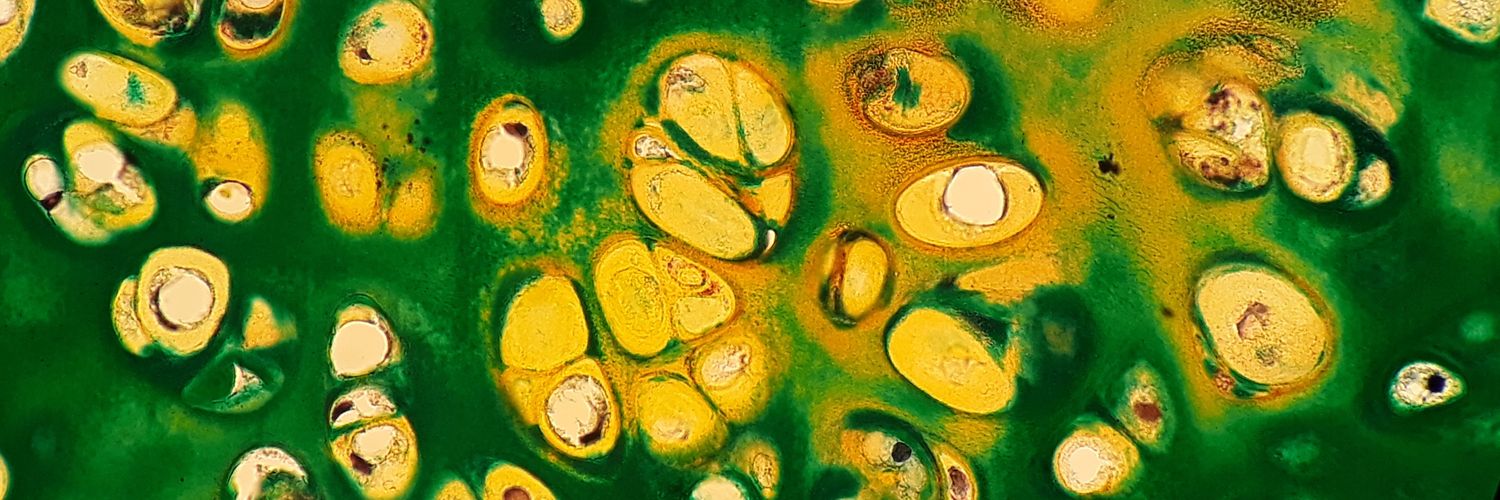
Just a gorgeous "Follicular Neoplasm" on thyroid FNA. As I often say, more important than the microfollicular pattern is the monotony of the follicles. Also seen here are some "flaming" cells (fire flames are by no means specific for Graves' disease).
Axillary lymph node. What is your diagnosis? 🔎 Stain clues and poll below 🗳️👇 #PathPuzzle 🕵️♂️ @KMirza @UMichPath #Pathology #PathX #PathTwitter
Spring 2025 Primary exam results have been released. Log into PATHway and visit the Board Correspondence page to see your results. abpath.org
This hepatic explant shows cirrhosis. The etiology is apparent on the PAS/D stain - alpha 1 anti-trypsin deficiency.
Interesting case to share (Board question): What's your diagnosis for this 3.3 cm renal tumor from a 42-year-old man? Vimentin, SMA, CD34, and CD10 are positive. Electron microscopy reveals electron-dense, rhomboid-shaped crystals in the cytoplasm.
50s M 4cm thyroid nodule. Dx/workup? #ENTpath #HeadandNeckPath #PathX #PathTwitter
Solitary breast mass in a 65 year old female, let me know your thoughts before I give the diagnosis :) #breastpath #pathtwitter #pathx #pathology #pathresidents
🔬💉Lymph node #FNA - malignant melanoma cells with enlarged nuclei, binucleation, and black pigment. Summer is here☀️It’s time to enjoy and protect yourself and your loved ones⚠️ 🧴⛱️🕶️ #cytopath #cytology