Dan Graham
@DanGrahamMD
General Pathologist at Clinical Labs of Hawaii / Interest in BST & H&N
Ectomesenchymal Chondromyxoid Tumor Synonyms: Oral myoepithelioma of soft tissue origin; reticulated myxoid tumor of the 👅; myoepithelioma of the 👅 Benign mesenchymal neoplasm of unknown histogenesis characterized by spindle-polygonal cells within myxoid-collagenous stroma &…
This is a GIST in the stomach. Most areas showed classic spindle-cell morphology w/ paranuclear vacuoles. Kudos @pepeheffernan , you got it!
Organ? Dx? #PathX #PathTwitter
BRG1, INI1 intact. NUT neg. Basaloid SCC as working dx
60s M neck and paraspinal masses. Bx of neck mass (imaging shown below). Dx/workup? #HeadandNeckPath #ENTPath #PathTwitter #PathX
#PathX #GlPath Rectum,polyp, Bx Dx: Rectum mucosa with infiltration by mantle cell lymphoma, classic type according to the WHO. Immunomorphologically, CD20+, Cyclin D1+, CD5 and CD3 are negative The proliferation rate (Ki-67) is ~10%.
Bladder involved by plasma cell myeloma #GUpath #Hempath ▶️Normal overlying urothelium ▶️Morphology varies from mature-appearing plasma cells (eccentric nuclei, spoke-wheel/clock-face chromatin) to immature, plasmablastic and pleomorphic variants (including multinucleated and…
MENINGIOMA, G1, parietal dura Interestingly, this psammomatous pattern was vast minority. Mostly fibrous. Focal meningothelial.
Organ? Dx? #PathX #PathTwitter
Dan, this focus is located within the renal parenchyma. There's another similar PNI on the same slide. My hypothesis is that the tumor's neurotrophic effect promotes nerve fiber growth, making them more prominent and easier to identify. See the text and figure below (PMID:…
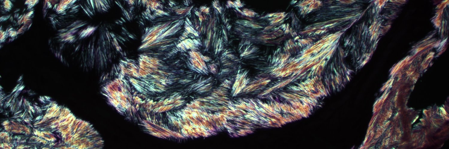
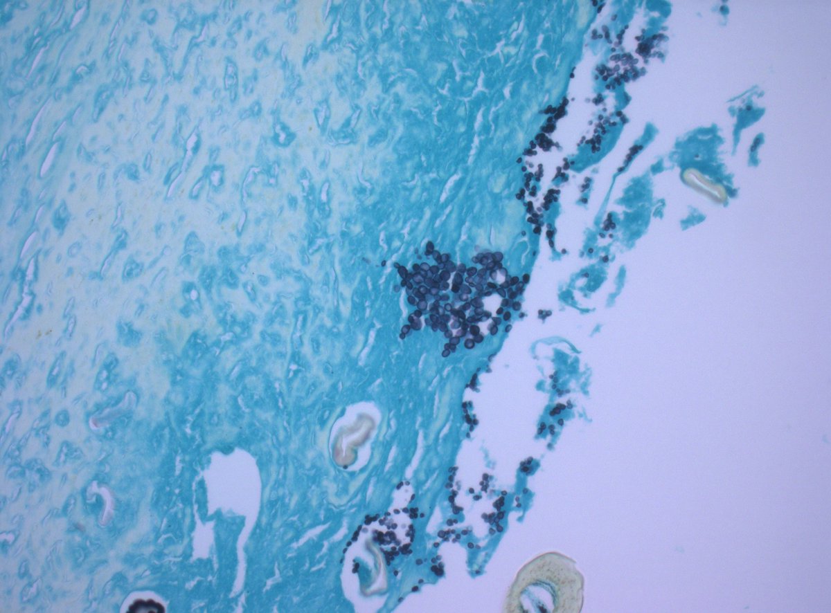
Seen in necrotizing granulation tissue in superficial arm mass of 20s F. Mysterious objects are lurking. Can you name them? #fungus #PathX #PathTwitter #pathbugs


So impressed you guys got this. INVERTED UROTHELIAL PAPILLOMA More pics below
Organ? Dx? #PathX #PathTwitter
😮 Very interesting Skin Tumor 🧂 Looks like Merkel. CK20 = perinuclear dot like. Neuroendocrine markers + 🧬 BUT CD99 diffusely membraneous and EWSR1-ERG fusion t(21;22) ---> (Primary) Cutaneous Ewing's! Open Access Article (Moubarak et al) ⬇️ doi.org/10.1111/cup.14…
This hepatic explant shows cirrhosis. The etiology is apparent on the PAS/D stain - alpha 1 anti-trypsin deficiency.

Interesting case to share (Board question): What's your diagnosis for this 3.3 cm renal tumor from a 42-year-old man? Vimentin, SMA, CD34, and CD10 are positive. Electron microscopy reveals electron-dense, rhomboid-shaped crystals in the cytoplasm.
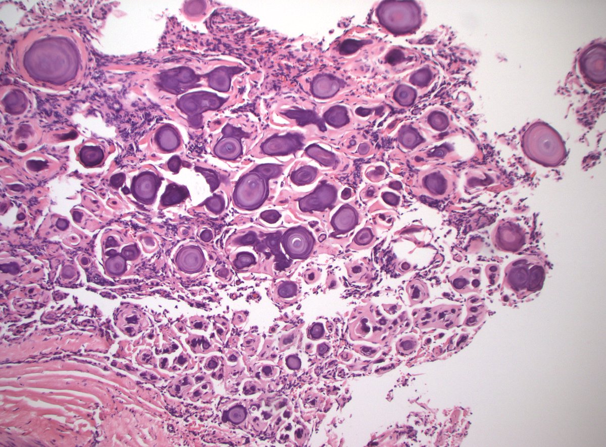
Foreign body reaction secondary to calcium hydroxyapatite cosmetic filler material
You all nailed it. Sinonasal papilloma, oncocytic type This case showed characteristic endophytic and exophytic growth, layered oncocytic cells with scattered mucocytes, microcysts, and neutrophilic microabscesses. The term Schneiderian membrane/epithelium/mucosa, and, by…
Organ? Dx? #PathX #PathTwitter

SF1, brachyury, DPC4, arginase1, BRG1, INI1 neg/normal MDM2 below. Thoughts? @HoustonArsenal @RibianszkyA
60s M retroperitoneal mass and inguinal mass/node. Bx from inguinal mass/node. CK+, p40/p63 -. Dx/further w/u? #PathX #PathTwitter