StainsFile
@StainsFile
Let’s talk about visualizing cells! 👩🏽🔬🔬 We’re an online resource for open-access protocols, reagents, theory, and more. Brought to you by @STEMCELLTech.
Welcome to StainsFile by @STEMCELLTech! Here, you can enjoy easy and open access to all the protocols and supporting resources you need to help you fix, prepare, and stain specimens: stainsfile.com 👩🏽🔬💻🧫🔬 #cellstaining #histology #microscopy #microscopymonday
Dr. Johann Danzl's team at @ISTAustria describe light-microscopy-based connectomics (LICONN) in their recent @Nature study. 🔬 LICONN allows for synapse-level phenotyping of brain tissue in biological experiments in a readily adoptable manner: go.nature.com/3H9WZva

🎧 How are changes in diagnostics affecting labs? Tune in to the On the Microscope Podcast from the @MayoClinic, where experts break down the biggest shifts in diagnostics and how they’re shaping clinical decision-making. 🎙️ apple.co/44owX0l #SciencePodcast
Our next episode features Dr. James Briscoe (@briscoejames) from @TheCrick! Check out his team's @Nature paper developing a 3D model of human trunk formation: go.nature.com/4lzKF77
PD-L1 IHC assays are FDA approved companion diagnostics that are critical to determine the PD-L1 expression levels in different tumors. 🖊️ Sign up for this @NS4Histotech webinar to hear recent updates about these assays: bit.ly/460HsZl

Need help with staining your epithelial organoids? Here's a protocol for immunostaining epithelial organoids from different tissues—including intestinal, mammary, prostate, lung, and pancreatic—after fixation: bit.ly/4nl9Uv3

🚨 Add some more color to your live-cell, super-resolution, and electron #microscopy! Researchers from Fujian Medical University have developed an extremely stable monomeric RFP named mScarlet3-H. Learn more in @naturemethods: go.nature.com/3RWW3MP

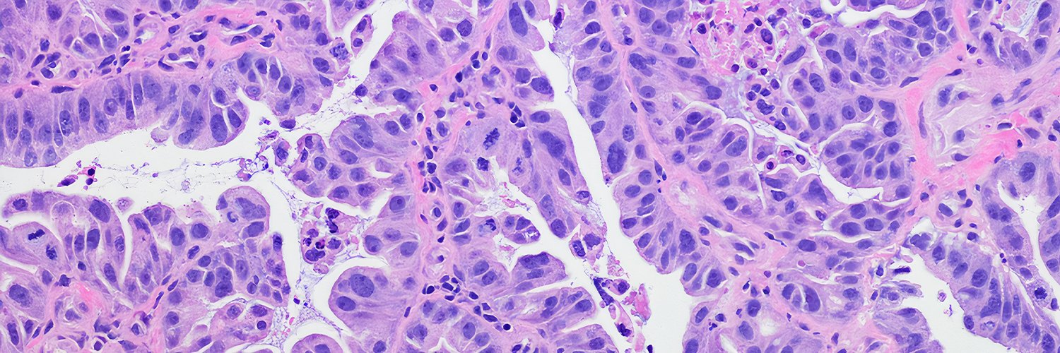
We would wonder where this hepatocellular carcinoma with large nuclei would lie in the model reported this month: Patil A, Hasan B, Park BU, Smith L, Sivasubramaniam P, Elhalaby R, Elessawy N, Nazli S, DaCosta A, Shabaan A, Cannon A, Lau C, Hartley CP, Graham RP, Moreira RK. A…
Case of Tubular Adenoma of Breast🔬 Tubular adenoma of breast is a benign mass composed of densely-packed round ductules with limited intervening stroma. No relation to the colon! #PathX #PathTwitter #breastpath
🫁 Pleural effusion. 🔬 Negative. Category 2 The International System for Reporting Serous Fluid Cytopathology. 🪟 Reactive changes. Windows. #Cytology #CytoPath #PathX
New in #HumPathol: Biliary adenofibroma and cholangiocarcinoma: neighbors or relatives? A systematic and critical review. sciencedirect.com/science/articl… #pathology #PathTwitter #PathX #liverpath
🔹Resinous versus aqueous mounting media? 🔹How do you use refractive index and diffraction to make your images look their best? 🔹What's ringing media? Mounting specimens doesn't have to be insurMOUNTable. 😉 Read up on mounting media in #histology: bit.ly/45zeoIa

Happy Friday! 😁🧫 When you study thousands upon thousands of cells, you're bound to find some patterns that make you smile. This image features a cell line that illuminates lamin B1, a protein in the nuclear envelope. #SciShots
While 3D cell cultures can more accurately represent complex tissues, what's the best way to image them? Dr. Jonathan Liu's (@jonliu123) team at @UWBioE reviews both 2D and 3D #microscopy approaches for studying 3D cell cultures. @naturemethods: go.nature.com/4efXv7F


New in #HumPathol: Triple-Negative Lobular Breast Cancer: focus on pathology and clinical challenges. sciencedirect.com/science/articl… #pathology #PathTwitter #PathX #breastpath
🔬 The @AllenInstitute has launched OpenScope, a groundbreaking platform that gives researchers around the world access to powerful brain imaging tools. 🧠 See how you can propose an experiment—at no cost—in the Allen Brain Observatory: bit.ly/3HOMzS4 #OpenScience

New in #HumPathol: BSND: An emerging immunohistochemical marker that reliably distinguishes benign from malignant oncocytic salivary gland tumors. sciencedirect.com/science/articl… #pathology #PathTwitter #PathX #ENTpath
Do you have beautiful images of stained or dyed specimens that you’d like to share with the scientific community and beyond? 🖼️ Submit your image to StainsFile and contribute to a growing, open-access histology resource! bit.ly/3TwzWxB

🔬👨💻📰 #SReD is out! Automated structural detection for #ImageJ & #FIJI, from nano to macro ✨🐘. No training data, no bias - texture analysis with GPU acceleration!⚡️ Brainchild of @afonsomendes92 and adventure w @christlet lab + friends. Check: nature.com/articles/s4146…
New research shows nonlinear live-cell imaging is a powerful tool for studying myelin morphology in conditions like #MultipleSclerosis. 🧠 Drs. Marie Louise Groot and @ALuchicchi at @amsterdamumc show subtle white matter changes in neurological donors. bit.ly/4ilrk7a

How do cells clean up misfolded proteins when they divide? This study finds that the cleanup crew includes the ER chaperone BiP and the proteasome, and kicks in right as cells finish dividing. Surprisingly, one major cell cycle regulator isn’t involved. elifesciences.org/articles/96675…