Ziad El-Zaatari
@ziad_zaatari
Surgical Pathologist @MethodistHosp. #GUpath #RenalPath. Photography and #PathArt enthusiast. Tweets do not equal medical advice.
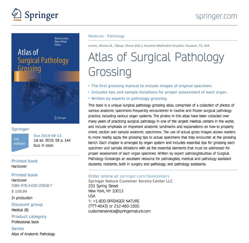
New Path Residents: Check out this Grossing Atlas with nearly 150 real gross specimens, sample dictations and tips. Written by Monica Lemos, an experienced PA and teacher at @MethodistHosp Book will be out in August, and you can pre order from Springer or Amazon. #grosspath
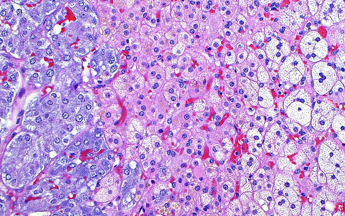
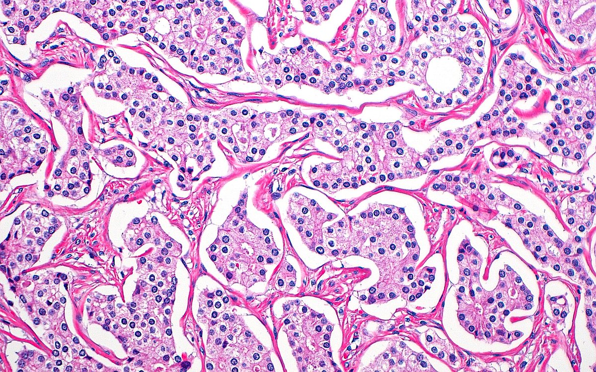
🔬 Pink, White and Blue 🔴⚪️🔵 ~ Adrenal cells at the intersection of cortex (right side) and medulla (left side) ~ #Histology #Pathology #GUpath #PathArt
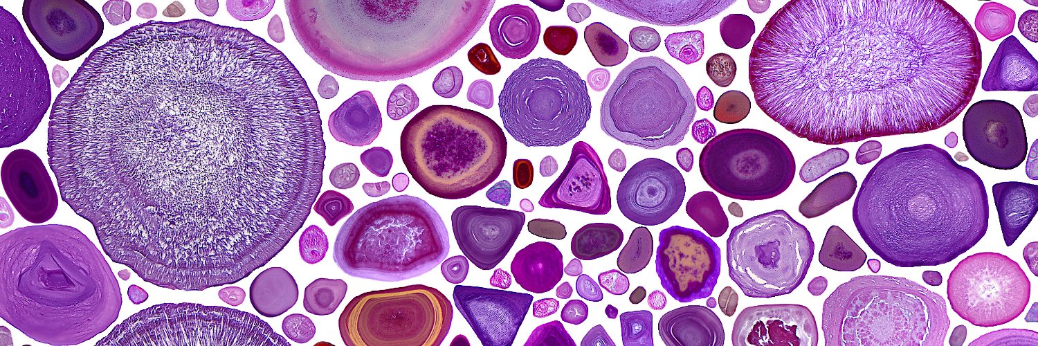
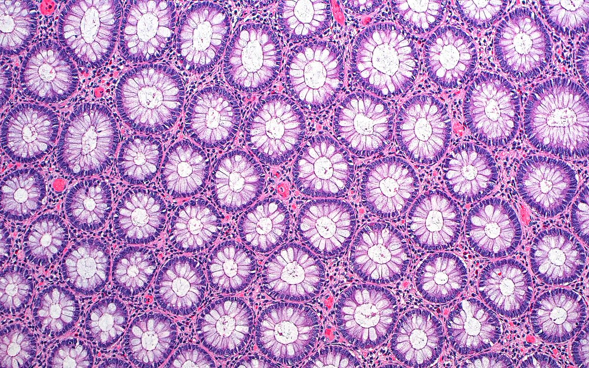
🔬📷🎨 Classic, basic, and beautiful...and never fails at making me stop to take a photo! ~ Normal Colonic Crypt Cross Sections: "A Sea of Flowers" 🌼🌸 #Pathology #Histology #PathArt
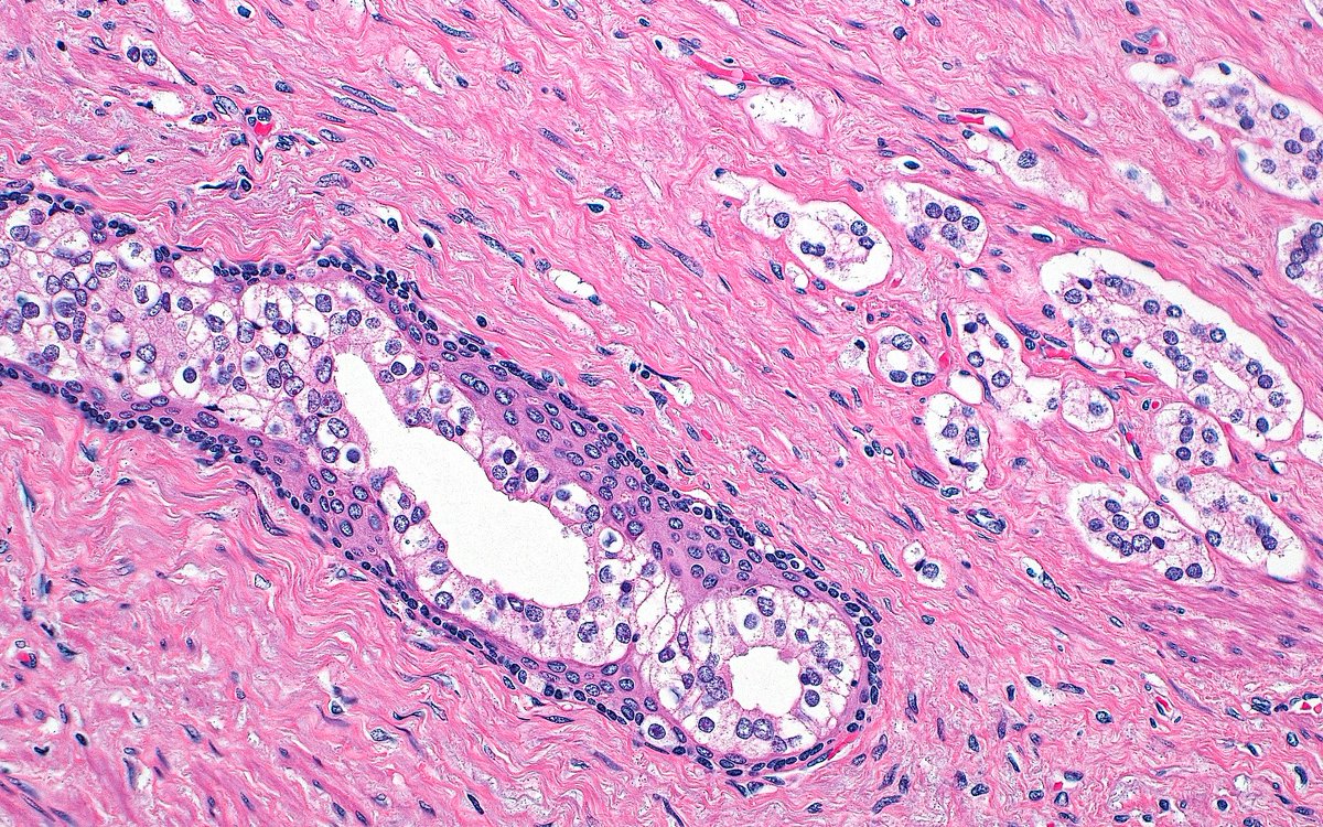
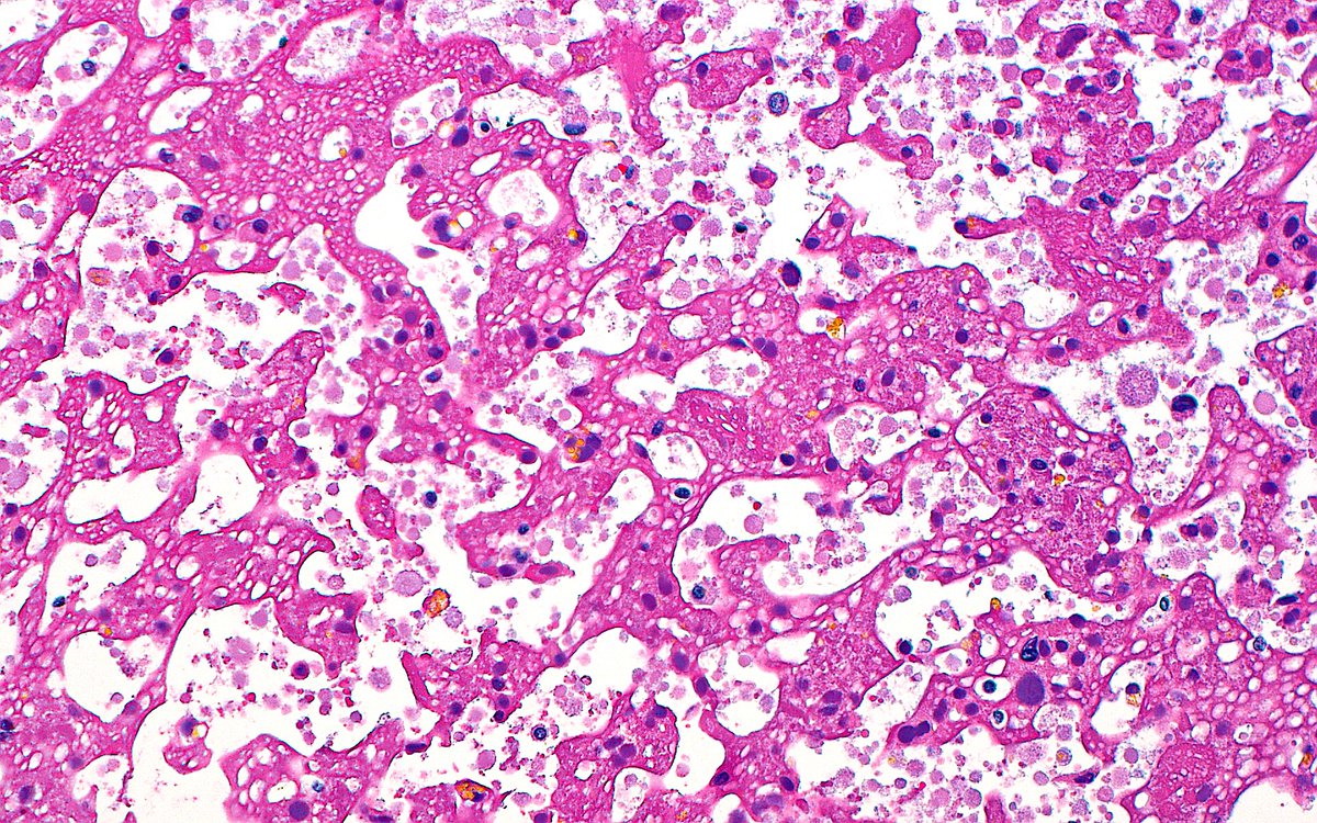
🔬📷 Seminal Vesicle Secretions ~ #GUpath #Pathology #Histology #PathArt 🎨
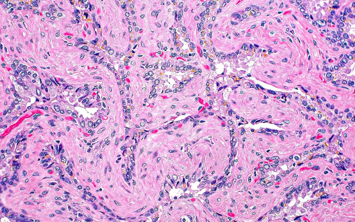
🔬 Prostate Cancer "Puzzle" 🧩 ~ Prostatic adenocarcinoma glands with retraction artifact ~ #GUpath #Pathology #PathArt
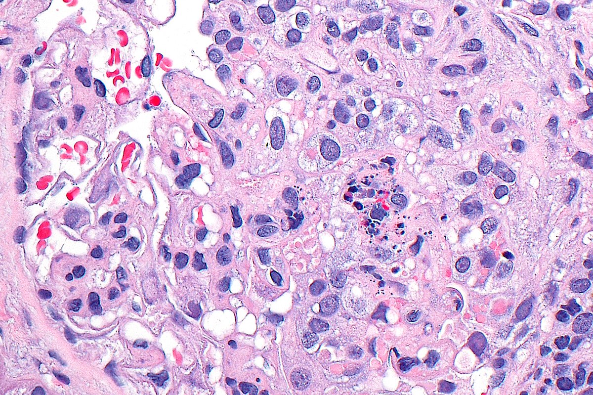
🔬📷 Glomerulus with karyorrhexis, hyaline thrombi, and a cellular crescent in patient with class III lupus nephritis ~ #RenalPath #Pathology #Kidney #Nephrology
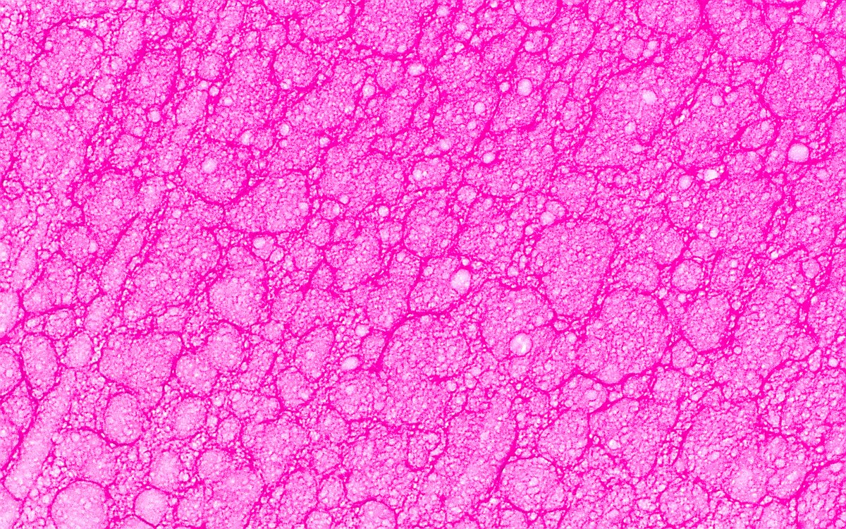
🔬 Cool Bubbles in Small Renal Cyst ~ #Pathology #Histology #RenalPath #SciArt #BioArt #PathArt 📸🎨
Colon histology with Melanosis coli. In watercolor. #pathart #pathology #pathx

The kidney, watercolor, 9x12” whitecoatartistry.etsy.com #PathArt #WhiteCoatArtistry
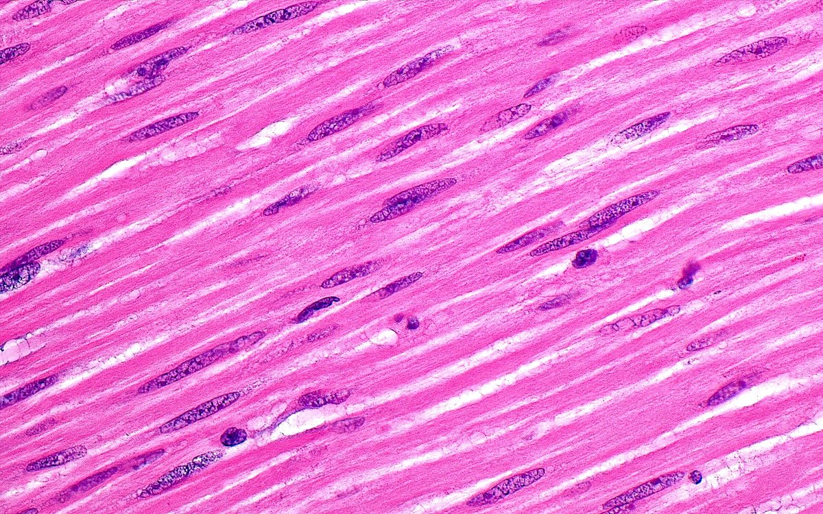
🔬 Smooth Muscle Cells ~ #Histology #Pathology #PathArt 📸🎨
🔬 Artery in a Kidney, Trichrome Stain ~ #Histology #Pathology #RenalPath #PathArt 🎨📸
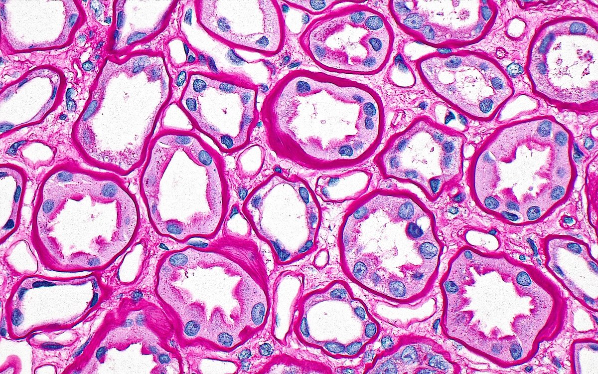
🔬 Proximal Renal Tubules (PAS stain) ~ #RenalPath #Histology #Kidney #Pathology #PathArt 🎨📸

June's image of the month highlights the beauty in cellular analysis 🔬 This artwork come from Selamete Çeku, who says “My #pathart reflects the essence of the pathologist’s profession – a commitment to transforming the future of healthcare.” Read more! ow.ly/Rqnj50Wg2hk
🔬 Seminal Vesicle Epithelium ~ #GUpath #Histology #Pathology #PathArt 📸🎨
🔬 Renal Tubules and Peritubular Capillaries (PAS Stain) ~ #RenalPath #Histology #Pathology #PathArt 📸🎨
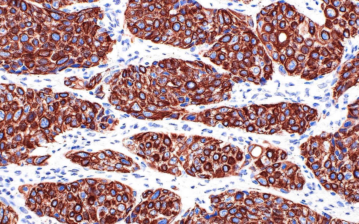
🔬 Invasive Urothelial Carcinoma, High Molecular Weight Cytokeratin Immunostain ~ #Pathology #IHCpath #GUpath #Histology #PathArt
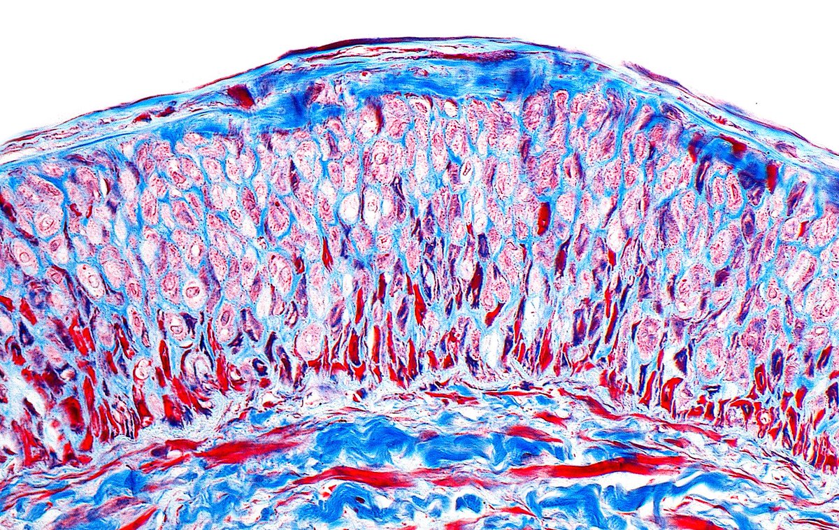
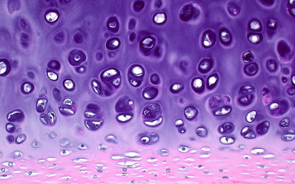
🔬 The Beauty of Cartilage ~ Hyaline cartilage from a the wall of a trachea ~ #Pathology #Histology #PathArt 🎨📸
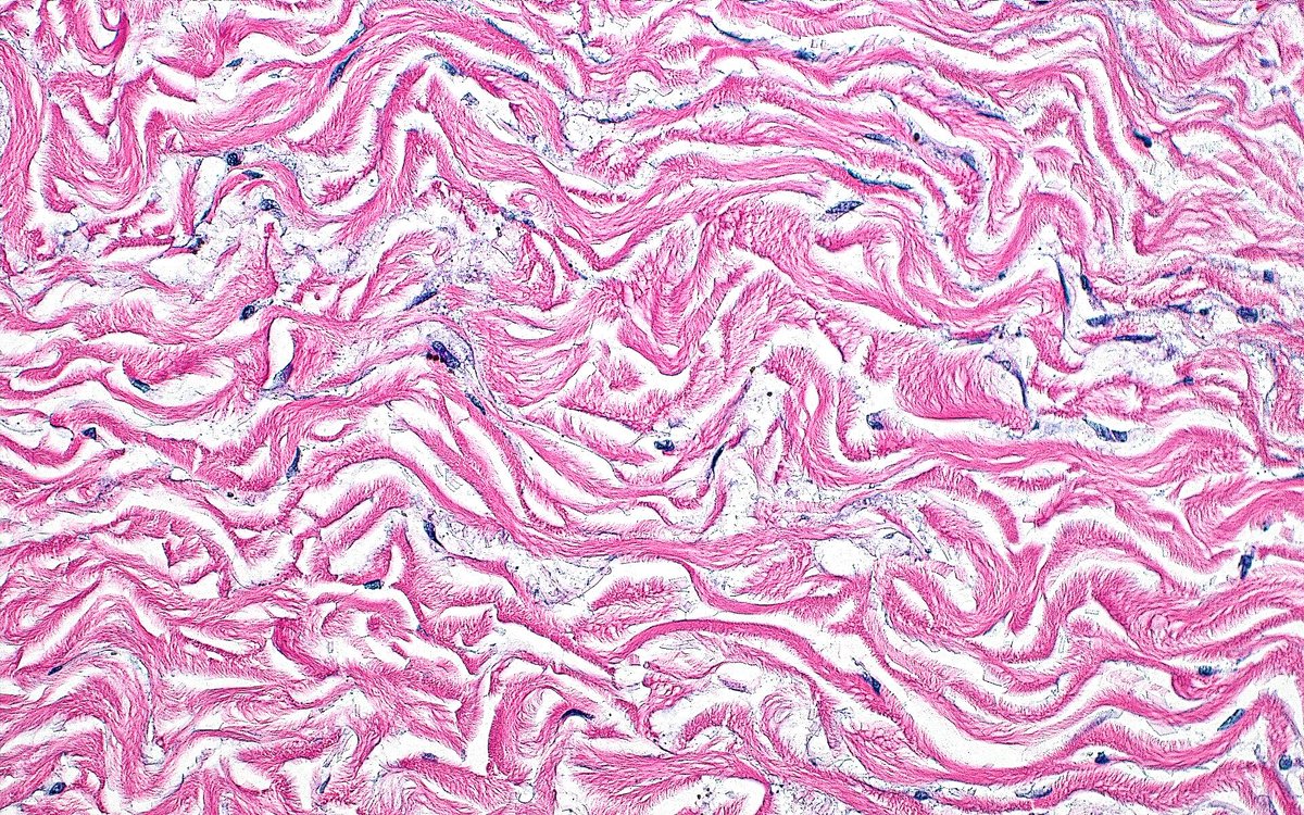
🔬 Collagen Fibers ~ From the adventitia of the aorta ~ #PathArt 🎨📸
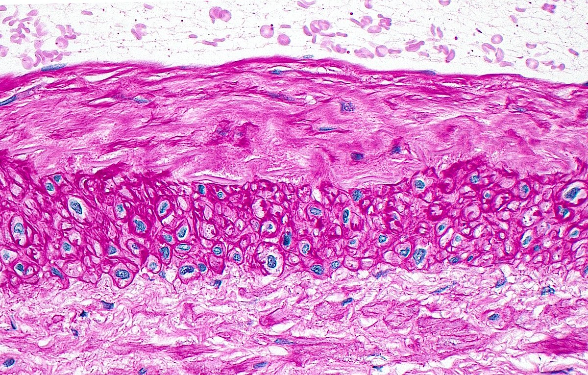
🔬 Layers of a Renal Artery (PAS Stain) ~ #Histology #Pathology #renalpath (From top to bottom): ▪️Lumen (containing RBCs) ▪️Intima (abnormally thickened here) ▪️Media (smooth muscle cells) ▪️Adventitia (outer connective tissue)
🔬 Intraductal Prostatic Carcinoma ~ Prostate cancer cells colonizing a benign prostatic duct (left) adjacent to high grade invasive prostatic adenocarcinoma glands (right) ~ #GUpath #Pathology #Prostate