Jack Garnham
@jjgarnham
Consultant neuroradiologist @ImperialNHS. Previous pan-London neuroradiology fellow. Winner of the Frank Doyle medal. Interest in medical education.
Be careful when interpreting trauma scans in patients with rigid spines. This unstable spinal hyperextension injury through the C4 - C5 intervertebral disc space (AOS B3) and facet joint injury (AOS F4) were sustained after a fall from sitting! Patients with rigid spines (e.g.,…


Terrible spino-pelvic injury after a three-story fall. This is an AOS C3, a displaced U-type sacral fracture. The fracture is complex and comminuted, but the morphology is in essence 1. bilateral craniocaudal fracture lines extending through the sacral foramina, and 2. a…


Bilateral medial medullary infarction (BMMI), resembling a heart (or a pair of AirPods, for the technologically inclined). At this level the medial medullary territory is supplied by branches from the anterior spinal artery. Bilateral infarction can occur with occlusion of a…


Multicentric glioblastoma with spectroscopy. Separate areas of cortical - subcortical thickening and FLAIR hyperintensity, with some areas of irregular parenchymal enhancement. Long TE single voxel spectroscopy demonstrates reversal of Hunter's angle, with decreased…


Beautiful hypertrophic polyneuropathy. A branch of L4 the nerve and the L5 nerve run along the anterior aspect of the sacral ala; these are massively enlarged. Can you also appreciate how big the exiting lumbosacral nerves are on the axial and sagittal images? The two most common…


Where have the parotid glands gone? This is parotid lipomatosis or liposubstitution! This particular case was secondary to previous treatment with radioiodine, which concentrates in the salivary glands, but it can also be seen as a consequence of normal ageing, autoimmune disease…


Elderly patient with a trans-spatial enhancing orbital mass with intraconal and extraconal components, squeezing around the optic nerve and extraocular muscles but respecting the confines of the bony orbit. These are the typical appearances of an orbital lymphoproliferative…


Opening the stack and scrolling down, you're confronted with this... You should almost always expect to see some sulci at the vertex, but you don't in this case. This is a portent of badness, so we're worried before we've even seen the rest of the study. Scrolling further down,…


This patient presented with a classic aneurysmal subarachnoid haemorrhage. We couldn't find an aneurysm on CTA, MRA, or DSA... but on a subsequent black-blood MRA there was a tiny fleck of enhancement in the region of the ACOM. On repeat DSA, this turned out to be a tiny…


Well-defined, ovoid, predominantly intraconal mass in the right orbit, displacing the optic nerve and extraocular muscles. T2 hyperintense with dynamic enhancement, patchy and heterogeneous at first with progressive filling on the delayed images These are the typical features of…


On a CT neck you see thickening and medialisation of the left aryepiglottic fold, an enlarged left pyriform sinus, and enlargement of the left laryngeal ventricle. What does this mean and what do you need to do next? The next step is a CT chest. These features suggest a left…


A young woman presents with acute, painful, monocular left-sided visual loss, worsening over a few days. On MRI we see enlargement, T2 hyperintensity, and enhancement of the intraorbital segment of the left optic nerve. This was acute optic neuritis. In adults, approximately 30%…


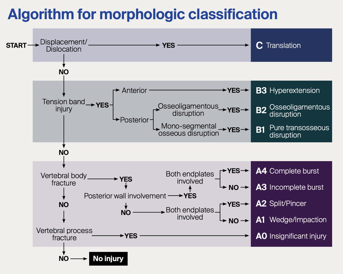
This T7 fracture involves two columns but is it unstable? The Denis three column model would suggest it is, but the reality is more complex. The three column model is now outdated. It doesn’t adequately account for soft tissue injuries, dynamic instability, or neurologic injury,…


How do you explain this tiny focus of diffusion restriction in the body of the right hippocampus? The patient was a confused adult with profound anterograde amnesia. This was transient global amnesia! TGA is a rare, benign, self-limited disorder characterised by failure of…


You could almost walk past this pre-medullary mass on T2 or FLAIR (is it just flow artefact?) but the DWI hyperintensity is striking and there is positive mass effect. This is an epidermoid cyst, a subtype of ectodermal inclusion cyst. These aren't neoplastic; they arise from…


What's your differential for an intraventricular mass in this location? What do you think about this predominantly solid markedly FLAIR hyperintense lesion in the left lateral ventricle? It's non-enhancing and there's no internal haemorrhage or calcification. This turned out to…


Early Fahr's disease (primary familial brain calcification or PFBC) with calcification in the lentiform nuclei, globi pallidi, dentate nuclei, and corpus medullare. Typically presents with progressive dystonia, parkinsonism, and neuropsychiatric features in adulthood. Most Fahr's…


We are all familiar with cavernous malformations (CMs) and their typical "popcorn" appearance and haemorrhagic rim. These benign non-neoplastic vascular lesions are seen in approximately 0.5% of people. CMs are composed of dilated thin-walled capillaries with an endothelial…


