Dr. Ruby Peters
@Ruby_Peters_
Physics of life fellow at the University of Sheffield. Physicist by training, exploring all things actin! #Actincortex #Superresolution #ImageAnalysis
🚨We’re hiring! Passionate about #microscopy, #biophysics and #interdisciplinaryresearch? There's still time to apply for our EPSRC PhD project on how actin network architecture shapes the mechanical properties of cells at @sheffielduni Learn more/Apply: findaphd.com/phds/project/f…

Very proud of @BoyaZhang4 and this lovely first paper, first of many I am sure.
My first author paper is now online in @OpticaWorldwide ! Please allow me introduce Vortex Light Field Microscopy: a simple and efficient way of achieving 3D spectral single-molecule imaging. Comments and feedbacks are welcome! @CamMicroscopy @PDN_Cambridge @Cambridge_Uni
Could not be prouder of my dear friend on this fantastic achievement!
My first author paper is now online in @OpticaWorldwide ! Please allow me introduce Vortex Light Field Microscopy: a simple and efficient way of achieving 3D spectral single-molecule imaging. Comments and feedbacks are welcome! @CamMicroscopy @PDN_Cambridge @Cambridge_Uni
Incredibly excited to share that I have joined @sheffielduni as an Early Career Research Fellow in the Physics of Life. My lab will use and develop fancy microscopes to investigate all things actin. I will soon be recruiting a PhD student, if you are interested, get in touch!

📰🔬🧠 Come explore the future of #microscopy in our new @JofMicroscopy review. As we dive into the #AI-assisted era, we examine pioneering studies opening the door for increasingly smarter sample-aware bioimaging systems. Kudos to @ALeonorMorgado et al onlinelibrary.wiley.com/doi/10.1111/jm…
Interested in understanding how #MachineLearning can empower data-driven #microscopy strategies? Check out our review, where we go from the basic concepts to an overview of the pioneering works in this emerging field! Just out in Journal of Microscopy: doi.org/10.1111/jmi.13…
🚨 JOB CALL!!! 🚨 If you’re interested in neuronal cell biology, come and work with us to use custom imaging and omics tools to study organelle contact sites in neurons. Please RT friends‼️ Open calls for a @wellcometrust funded Post doc and RA position listed below…
Excited to finally share my first first-author paper out now in @NatureComms! 🔬 SMLFM offers a much-needed optical solution in 3D-SMLM, enabling a 10x speed advantage in single molecule imaging and tracking over large volumes! 🦠 nature.com/articles/s4146…
Another exciting paper from another awesome colleague this week - congrats @GeraldineJowett !
Interested in understanding how #MachineLearning can empower data-driven #microscopy strategies? Check out our review, where we go from the basic concepts to an overview of the pioneering works in this emerging field! Just out in Journal of Microscopy: doi.org/10.1111/jmi.13…
Great to see this out! Congratulations @NezaVadnjal and the rest of the team!
My first paper from my PhD with @PaluchLab at @LMCB_UCL and @PDN_Cambridge is out in @J_Cell_Sci: Proteomic analysis of the actin cortex in interphase and mitosis journals.biologists.com/jcs/article/13…
Could not be prouder of you @GriffieJuliette - one to watch for sure!
I am delighted to announce that I will be starting my own lab next year @scilifelab in Stockholm!! 🥳 [1/3]
You work with budding or fission yeast, and are interested in #superresolution ultrastructure imaging with conventional microscopes? We have the #UExM protocol for you! biorxiv.org/content/10.110… 1/2

Tired of counting cell divisions? Bored of manually detecting a specific event from your movies? Don't want to spend half your PhD clicking on cells 🙃? I spent half of mine for you: 👇🧵 biorxiv.org/content/10.110…
Could not be prouder of my colleague and dear friend who is going to be an amazing PI - when can I join ?! So happy to see you go onto the next adventure!
I am delighted to announce that I will be starting my own lab next year @scilifelab in Stockholm!! 🥳 [1/3]
😍
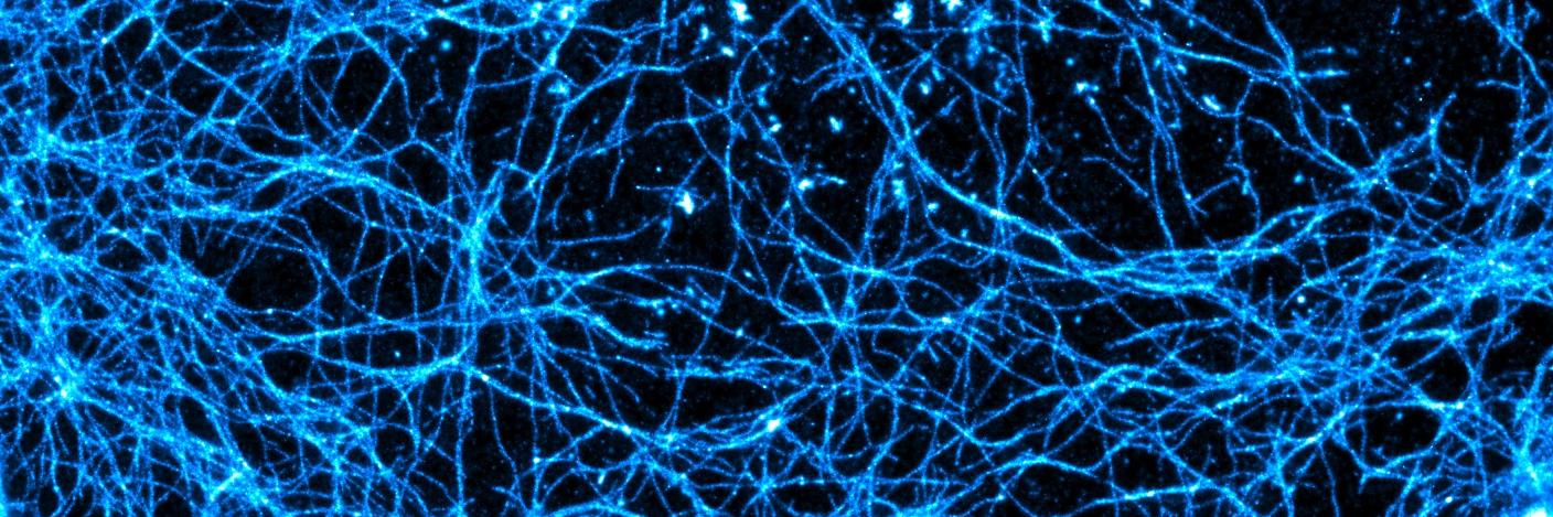
The #CytoMorphoLab is happy to present: Microtubule Mechano-Sensations, recorded for #cellbio2020 Feel free to comment the science or the movie and to share it with cell biology and cytoskeleton afficionados. vimeo.com/488648110
Best news!
The #CytoMorphoLab is happy to present: Microtubule Mechano-Sensations, recorded for #cellbio2020 Feel free to comment the science or the movie and to share it with cell biology and cytoskeleton afficionados. vimeo.com/488648110
Our latest paper in collab. with @Owen_lab_UoB, Duncan Gowland (@CANES_CDT) & Nicola Bonini is out. Adam Suhaj used fluorescence exp’ts & all-atom MD sims to investigate how Laurdan & di-4-ANEPPDHQ influence the properties of lipid membranes. Check it out: bit.ly/3n2agac
Microscopy Mavens! - Start spreading the news: 2x PDRA positions, ch.cam.ac.uk/job/27691 and a Research Assistant ch.cam.ac.uk/job/27693
Open postdoc position(s) available. Want to understand cell contractility, or just watch stuff move around inside cells? Joint lab space/meetings with @pwoakes means super-res + patterning + traction-force, etc. Please RT. DM or email. sites.google.com/view/jordanbea…